Translate this page into:
Multiple Cream-Colored Papules Over the Trunk and Neck
Address for correspondence: Dr. Vijay Shankar S, Department of Pathology, Adichunchanagiri Institute of Medical Sciences, BG Nagar - 571 448, Karnataka, India. E-mail: docpapu@gmail.com
This is an open-access article distributed under the terms of the Creative Commons Attribution License, which permits unrestricted use, distribution, and reproduction in any medium, provided the original work is properly cited.
This article was originally published by Medknow Publications and was migrated to Scientific Scholar after the change of Publisher.
A 55 year-old male presented with asymptomatic, tiny, raised lesions of six years' duration on the chest, abdomen, and back. These lesions had gradually increased in size and number. None of the family members were affected.
Examination revealed cream-colored, dome-shaped papules of 2-5 mm size over the root of the neck, chest [Figure 1], abdomen, back [Figure 2], and also on the medial arms. The lesions did not have any punctum. A small skin-colored papule of 2 mm size was present on the right scrotal wall. Nails were normal.

- Multiple cream-colored papules on the chest and neck

- There are few comedones in addition to the creamish papules on the back
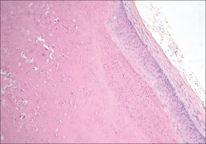
An excisional biopsy was performed from a papule from the back; the histological picture is seen in Figures 3 and 4.

- Collapsed cyst whose walls are thrown into folds and lined by squamous epithelium (H and E, ×40)

- Higher magnification of the cyst wall (H and E, ×400)
WHAT IS YOUR DIAGNOSIS?
DIAGNOSIS: STEATOCYSTOMA MULTIPLEX
Histology revealed a collapsed cyst in the dermis. The cyst wall was lined by multiple layers of stratified squamous epithelium [Figure 3]. Lobules of sebaceous glands could be seen in the wall of the cyst. The luminal surface of the cyst wall revealed homogeneous eosinophilic cuticle at foci [Figure 4].
DISCUSSION
Steatocystoma multiplex (sebocystomatosis) is an autosomal dominant condition[1] that usually presents in adolescence. It is characterized by multiple, yellowish or skin-colored, dome-shaped nodules and cysts over the trunk, proximal extremities, and the face. However, acral distribution has also been reported.[2] The common sites of involvement are the chest and the arms, but these nodules can occur anywhere in the body. Sometimes, the condition can manifest as a solitary lesion, when it is called steatocystoma simplex.
These papules have to be clinically differentiated from other cysts. Eruptive vellus hair cysts are commonly found on the chest. Vellus hair cysts are reddish brown in color and may be associated with itching. Epidermoid cysts and occasionally, vellus hair cysts may have a black punctum. Trichilemmal cysts are more common on the scalp.[34]
As the clinical features are variable, diagnosis may be difficult and require biopsy to be confirmed.
Steatocystoma multiplex and simplex arise from the ducts of sebaceous glands.[5] Histologically, these cysts are characterized by folded and crenated cyst walls that are lined by several layers of stratified squamous epithelium with wavy eosinophilic cuticles towards the luminal surface.[6] In atrophic areas, the lining may be one to three layers of flattened squamous epithelium. The diagnostic feature is the presence of lobules of sebaceous glands, which are either close to or seen within the lining epithelium. Occasionally, the lining of the cyst wall may contain macrophages.[1] Sabater-Marco et al. have reported the presence of smooth muscles in the cyst wall. [7] These cysts are usually empty as the predominantly lipid contents are washed away during the processing of the biopsy specimen. Uncommonly, homogeneous, light-staining, eosinophilic contents and vellus hairs may be visualized in the cavity of the cysts. Steatocystoma has to be differentiated histologically from trichilemmal cysts [Figure 5], eruptive vellus hair cysts [Figure 6], and infundibular cysts [Figure 7].

- Trichilemmal cyst contains homogenous, eosinophilic, compact, orthokeratotic material within the sac and the lining epithelium characteristically lacks granular layer

- Typical vellus hair cyst: Admixed with the lamellated keratin, there are numerous vellus hairs, which is very typical of vellus hair cyst

- Infundibular cyst: Cyst wall is composed of stratified squamous epithelium with granular layer. Note the lamellated keratin in the cavity
Table 1 gives the salient features of different types of cutaneous cysts.
| Cysts | Epidermoid/Sebaceous cyst | Trichilemmal/Pilar cyst | Steatocystoma | Eruptive vellus hair cyst |
|---|---|---|---|---|
| Age | Young and middle-aged | Young and middle-aged | Adolescence | Children and young adults |
| Distribution | Face, neck and upper trunk | Scalp | Trunk, upper extremities | Parasternal area |
| Morphology | Smooth dome-shaped swelling | Smooth dome-shaped swelling | Small dome-shaped translucent or yellowish | Soft, flesh-colored or reddish brown papules |
| Punctum | Present | Absent | Absent | May be present |
| Origin | Infundibulum of hair follicle | Isthmus of hair follicle | Duct of sebaceous glands | Arise in a developmental defect of hair follicle at the level of the infundibulum |
| Granular layer in the lining epithelium | Present | Absent | Absent | Present |
| Contents | Lamellated keratin | Homogeneous keratin | Most of the times, it is empty. If present, it is homogeneous and lightly stained. | Lamellated keratin with fine vellus hairs |
| Relation to the sebaceous glands | None | None | Lobules of sebaceous glands are seen within the lining epithelium or in close approximation to the cyst wall | None |
| Immunohistochemistry | Express K10 | Express K10 and K17 | Express K10 and K17 | Express K17 |
Steatocystoma is considered as forme fruste of pachyonychia congenita II and has been reported to be associated with mutations of keratin 17 gene in familial disease cases.[8‐10] Steatocystoma has been found in association with various conditions like multiple keratoacanthomas, rheumatoid arthritis, hyperkeratotic lichen planus, hidradenitis suppurativa, acrokeratosis verruciformis of Hopf, ichthyosis, koilonychias, and bilateral preauricular sinuses.[1112]
Various modalities of treatment such as surgery, laser, and cryotherapy have been employed in treating this condition. Most of the surgical techniques involve excision of the cyst in toto or decapitation of the cyst, extirpation of its contents, and removal of the cyst wall. Cysts are opened by perforating the cysts with a needle tip, or by stab incision with a surgical blade no. 11, or by electrocautery, or by CO2 laser. [13‐18] The contents are then expressed out and the cyst wall removed or destroyed competely to prevent recurrence. Cyst walls can also be destroyed chemically or by CO2 laser or electrosurgery.[16] Isotretinoin (1 mg/kg/day) has been used with variable results in extensive steatocystomas.[1920]
Source of Support: Nil
Conflict of Interest: None declared.
REFERENCES
- Steatocystoma multiplex: A case with unusual clinical and histological manifestation. Am J Dermatopathol. 1997;19:89-92.
- [Google Scholar]
- Disorders of sebaceous glands. In: Burns T, Breathnach S, Cox N, Griffiths C, eds. Rook's Textbook of Dermatology (7th ed). Massachusetts: Blackwell Science Publishing; p. :43.1-43.74.
- [Google Scholar]
- Tumors and cysts of epidermis. In: Elder D, Elenitsas R, Jaworsky C, Johnson, eds. Lever's Histopathology of the Skin (9th ed). Philadelphia: Lippincott Williams and Wilkins; 2005. p. :805-866.
- [Google Scholar]
- Tumors and related lesions of sebaceous glands. In: Mckee PH, Calonje E, Granter SR, eds. Pathology of skin withclinical correlations (3rd ed). Philadelphia: Elsevier Mosby; 2005. p. :1572-4.
- [Google Scholar]
- Simultaneous occurrence of multiple trichoblastomas and steatocystoma multiplex. Am J Dermatopathol. 1997;19:294-8.
- [Google Scholar]
- Steatocystoma multiplex with smooth muscle: A hamartoma of the pilosebaceous apparatus. Am J Dermatopathol. 1996;18:548-50.
- [Google Scholar]
- Expression of keratins (K 10 and K 17) in steatocystoma multiplex, eruptive vellus hair cysts, and epidermoid and trichilemmal cysts. Am J Dermatopathol. 1997;19:250-3.
- [Google Scholar]
- Steatocystoma multiplex. Created on 2nd June 1986. Available from: http://www.ncbi.nlm.nih.gov/entrez/dispomim.cgi?id=184500 [updated on 1998 Jul 2]. [accessed on 2008 Nov 24]
- [Google Scholar]
- Keratin 17 mutations cause either steatocystoma multiplex or pachonychia congenital type 2. Br J Dermatol. 1998;139:475.
- [Google Scholar]
- Steatocystoma multiplex with bilateral preauricular sinuses in four generations. Ann Plast Surg. 1988;21:55-7.
- [Google Scholar]
- Perforation and extirpation of steatocystoma multiplex. Int J Dermatol. 2009;48:329-30.
- [Google Scholar]
- A simple surgical technique for the treatment of steatocystoma multiplex. Int J Dermatol. 2001;40:785-8.
- [Google Scholar]
- Steatocystoma multiplex, treatment with electrosurgery: A novel approach. Indian J Dermatol. 1998;43:148-51.
- [Google Scholar]
- Late onset of a facial variant of steatocystoma multiplex - calretinin as a specific marker of the follicular companion cell layer. J Dtsch Dermatol Ges. 2008;6:480-2.
- [Google Scholar]
- CO2 laser therapy in a case of steatocystoma multiplex with prominent nodules on the face and neck. Int J Dermatol. 2003;42:302-4.
- [Google Scholar]
- Steatocystoma multiplex suppurativum: Oral isotretinoin treatment combined with cryotherapy. Australas J Dermatol. 2000;41:98-100.
- [Google Scholar]
- Isotretinoin in the treatment of steatocystoma multiplex: A possible adverse reaction. Cutis. 1986;37:115-20.
- [Google Scholar]


