Translate this page into:
Structural Changes of Fat Tissue After Nonaspirative Ultrasonic Hydrolipoclasy
Address for correspondence: Dr. Belchiolina B Fonseca, Biomedical Sciences Institute, Av. Pará, 1720, Campus Umuarama, CEP 38400-902, Uberlândia-MG, Brazil. E-mail: bialucas@yahoo.com.br
This is an open-access article distributed under the terms of the Creative Commons Attribution-Noncommercial-Share Alike 3.0 Unported, which permits unrestricted use, distribution, and reproduction in any medium, provided the original work is properly cited.
This article was originally published by Medknow Publications and was migrated to Scientific Scholar after the change of Publisher.
Abstract
Background:
Hydrolipoclasy is an alternative technique less invasive than liposuction. Hydrolipoclasy uses normal saline or hypotonic solution and ultrasound waves that act directly on local adiposity. In the literature there are few reports of morphostructural changes on adipose tissue.
Materials and Methods:
This study was aimed to evaluate the amount of fat cells injured immediately after treatment and after three days and also cell migration to the area treated using 8 pigs as experimental models, as well as cellular changes by transmission electron microscopy (TEM).
Results:
The Wilcoxon test was conducted, and a difference was found between the treated side and the corresponding control side on the number of viable cells. The treated side showed a smaller number of viable cells compared to the control side both immediately after treatment and 3 days later. Also occurring 3 days after treatment was the migration of lymphoid cells and fibroblasts, which shows the local inflammatory process and conjunctive neoformation. Soon after treatment there was fluid accumulation within adipocytes.
Conclusions:
The results shown in this paper demonstrate Ultrasonic Hydrolipoclasy as a viable alternative for the treatment of localized fat deposits without the side effects of traditional surgical procedures. Better results are expected when hypotonic solution is used, since it penetrates into the cell.
Keywords
Adipose tissue
alternative technique
ultrasound
INTRODUCTION
Ultrasonic hydrolipoclasy (HLC) was officially proposed in 1990 in Italy during the national congress of the International Society of Aesthetic Medicine and in 1991 at the World Congress of Aesthetic Medicine in Rio de Janeiro by Italian physician Ceccarelli. This technique consists of applying a hypotonic solution directly into the fatty tissue and then subjecting the tissue to the application of 3 MHz ultrasound. This makes the already swollen adipocytes become more susceptible to the action of ultrasonic waves; and, due to the phenomenon of cavitation, “explode,” releasing fat from within. These will be removed from the site via the lymphatic system.[12]
Doctors worldwide have been using HLC successfully, but the experiments are still restricted to few centers, such as those in Rome, Italy, by Ceccarelli. The research conducted on the microstructural changes of adipose tissue using the HLC, however, are not always easy since there is a need to use animals as experimental models. Mice are not suitable models, because they are very small and would probably not withstand the effects of ultrasound. An animal that could be used as a model would be the pig, due to its size and morphofunctional similarities with the human species.
Due to the importance of esthetic medicine in the world, the frequent demand of alternatives for control of local adiposity and the scarcity of scientific studies with experimental models, this study aims to assess the morphological and microstructural changes of adipocytes caused by using ultrasonic hydrolipoclasy upon Landrace pigs, which have thin adipose tissue, to better understand the effectiveness of this technique in humans.
MATERIALS AND METHODS
Eight 90-day-old female Landrace pigs, weighing 75 kg on average with about 2 cm of adipose tissue in the abdominal region were used. The animals were raised on a farm located in the industrial region of the Triângulo Mineiro, Brazil, where the HLC and the removal of tissue were performed.
For containment, procedure, and sample collection, animals were anesthetized with tiletamine associated to Zoletil intramuscularly. After restraint, a midline abdominal division was drawn (corresponding topographically to the ventral midline) and, using a tape measure, points were predefined and scored for completion of hydrolipoclasy and collection of tissues to be analyzed. The left side was predetermined as the test side and the contralateral (right) as the control side. On the test side, after cleansing with iodine alcohol, 120 ml of hypotonic solution (distilled water) was injected with a 30 G 1/2 hypodermic needle and the area was subsequently treated with ultrasound in continuous mode in the protocol for hydrolipoclasy, with 3 MHz ultrasound, at a power of 1.9 Watts/cm2 for 30 mins. After this period, with the same equipment used in hydrolipoclasy, lymphatic drainage was performed with ultrasound in pulsed mode at 50% power (1 Watt/cm2) for 10 mins.
On the control side no substance was injected nor any treatment performed with the device. Soon after the procedure (zero hour) and after 3 days skin fragments from the skin to the first layer of abdominal muscle were removed from both sides measuring approximately 2.5 cm3. Each fragment was divided into two, with one of the shares fixed in 10% formaldehyde in 0.1 M phosphate buffer, pH 7.2, to perform optical microscopy and the other part fixed in 3% glutaraldehyde in phosphate buffer at 0.1 M, pH 7.2, for transmission electron microscopy (TEM). The material was sent to the Laboratory of Histology and Electron Microscopy Center, Federal University of Uberlândia, for processing the material.
Optical microscopy
After 48 h of storage, samples were cut into smaller segments and washed in water, then dehydrated in increasing alcohol series to 50%, 70%, 85%, 90%, 95%, and 100% being 30 min at each concentration, washed in xylene (100%) and embedded in histological paraffin. Histological specimens 5–7 μm thick were stained with hematoxylin–eosin and mounted in Canada balsam.
The number of fat cells intact for μm2 was evaluated at zero hours and 3 days after treatment. To do this, digital images were captured on an Olympus BX40 binocular microscope with a 10× objective coupled to an Olympus OLY-200 camera that was connected to a PC via a Data Translation 3153 digitizer board. The images were evaluated using HL Image software. The counting was done in the first layer of fatty tissue underlying the skin and the result was an average of 8 different fields for each sample.
In addition to analyzing adipose cells three days after treatment, a count of cell nuclei by μm2 to check cell migration to repair tissue (lymphoid cells, fibroblasts, and adipocytes) was performed. The counting was automated using the same microscope and program described above, the result being the average of 8 different fields for each sample.
Electron microscopy
The fragments for TEM were cut into cubes of about one cubic millimeter and placed in a 1:1 solution of osmium tetroxide and 0.1 M phosphate buffer, pH 7.2, for 1 h and subsequently treated with potassium ferrocyanide (1.25%) for 30 mins. The fragments were dehydrated in increasing series of acetone. Subsequently, the material was placed in a 2:1 solution of Epon resin and propylene oxide for 12 h. After this period, the solution containing the material was placed in an oven at 37°C for 4 h and then the solution of resin and propylene oxide was replaced by a solution of pure resin in which the material was soaked for 4 h more. Finally, the cubes were embedded in Epon resin to be cut by ultramicrotome to obtain ultrathin cuts.[3]
The ultrathin sections were stained with uranyl acetate and subsequently with lead for 30 mins at room temperature.[3] The assessment and photographic documentation were made on an electron microscope (Zeiss Electron Microscope EM 109) and captured by MEGAVIEWG2 OLYMPUS SOFT IMAGE SOLUTION (Zeiss).
Ethics
We used the method of stunning indicated for swine and the method of euthanasia indicated by the law Conselho Federal de Medicina Veterinária, Brazil, number 714 from June 20, 2002.
Statistics
The statistical design was randomized using the nonparametric Wilcoxon test[4] with a significance level of P = 0.05. Analyses of variables were performed using the statistical program S-Plus 2000 (MathSoft, Inc.).
RESULTS
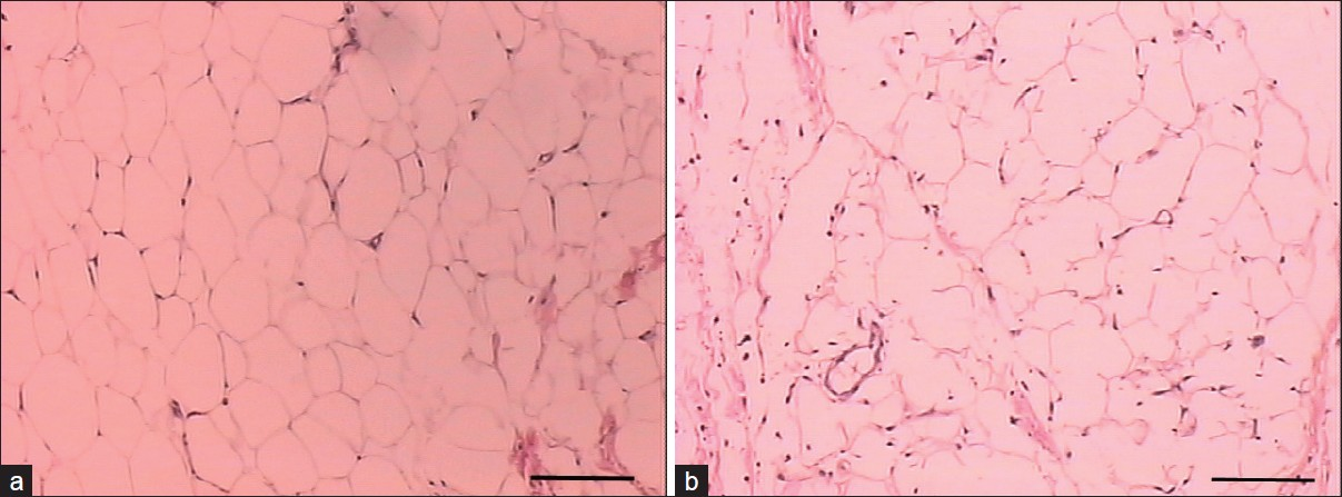
In light microscopy there was a difference between the amount of viable cells per field both immediately after the procedure and after 3 days of completion of HLC between test and control groups, whereas the test group showed a smaller number of cells per 100,000 μm2 (P < 0.05). Figure 1 shows pictures with the differences between test and control groups at zero hour; Figure 2, the difference 3 days after the procedure of HLC. Table 1 shows the number of cells per 100,000 μm2.

- (a) Pig fat tissue of the control side at zero hour (bar = 200 μm); (b) Pig fat tissue of test side after treatment with hydrolipoclasy (HLC) at zero hour (bar = 200 μm)

- (a) Pig fat tissue of the control side 3 days after treatment with HLP (bar = 200 μm); (b) Pig fat tissue of test side 3 days after treatment with HLP (bar = 200 μm)

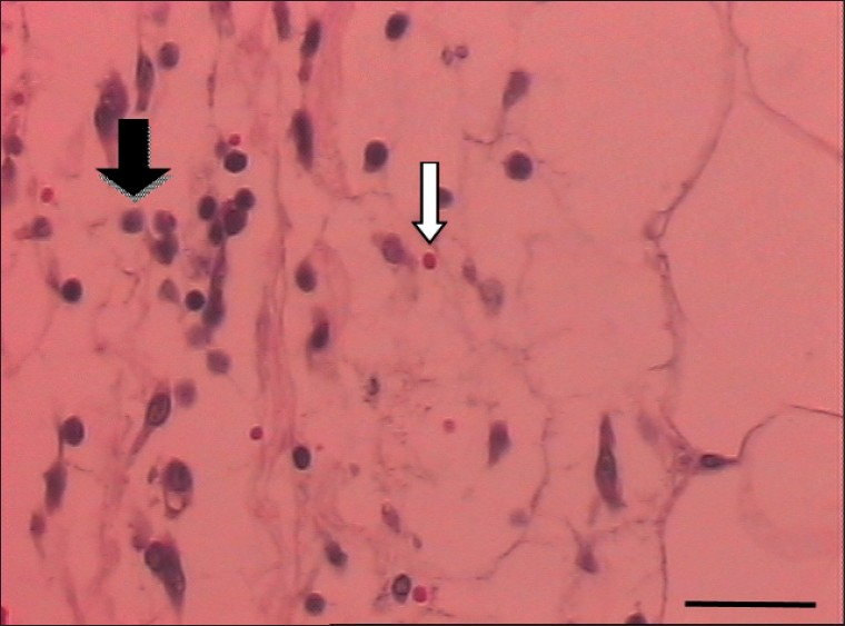
The test group had a higher number of migration cells per 10,000 μm2 in the control group 3 days after treatment. The last line of table 1 shows the number of cells per 10,000 μm2. Figure 3 shows that there was migration of lymphocytic cells and connective tissue formation, indicating a healing process.

- Cell migration in adipose tissue of pigs 3 days after treatment with HLP with a predominance of lymphocytes (black arrow). Red blood cells are also visible (white arrow) (bar = 50 μm)
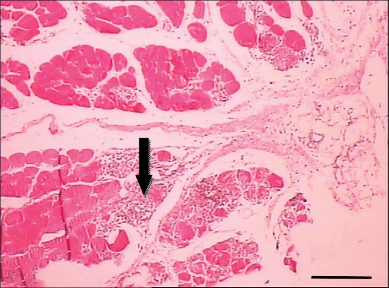
Another important finding in light microscopy is that the first animal, due to differences in skin thickness and consistency of human and pig tissues, suffered a puncture accident in infiltration with subsequent muscle necrosis and hemorrhage [Figure 4], showing the importance of the procedure to be performed by a physician knowledgeable in human anatomy.

- Areas of muscle necrosis 3 days after treatment with HLC, indicated by arrow, are caused by accidental puncture due to injection of the hypotonic solution into the skeletal muscle underlying the fatty tissue of a pig (bar = 500 μm)
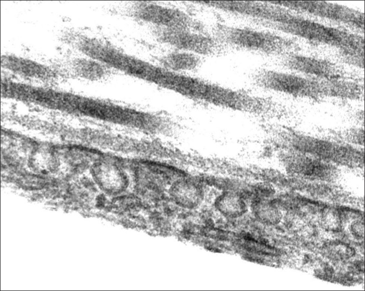
TEM showed that fluid had accumulated within the adipocytes of the test group immediately after the procedure at both superficial and deep levels [Figures 5a–c]. These accumulations were not found in the control group at any time, nor in the test group after 3 days of the experiment. Furthermore, the presence of vesicles in some micropinocitose adipocytes was shown [Figure 6].

- Areas of fluid accumulation within adipocytes immediately after the hydrolipoclasy procedure; (a) superficial portion (near the skin); (b) medium; and (c) deep portion (near the muscle)

- Formation of microvesicles of pinocytosis in adipocyti membrane
DISCUSSION
The differences between the number of viable cells presented between test and control groups in OM [Figures 1 and 2, Table 1] shows that hydrolipoclasy is an effective procedure for local adiposity. For over 30 years, ultrasound has been widely used by medical professionals.[5] The frequencies of the transducers play an important role in the treatment. Ultrasound with a frequency of 1 MHz is absorbed by tissues at depths between 3 and 5 cm. Longer treatments with a frequency of 3 MHz can promote therapeutic effects at depths between 1 and 3 cm.[6–8]
Knowledge and proper use of ultrasound is essential to achieve the best result of the technique, so the physician should be responsible for stating power, frequency and length of use of ultrasound in each case. This is necessary because low doses of ultrasound do not have the capacity to promote dissolution in fat cells. In general, the frequency of the transducer used in cosmetic procedures is equal to 3 MHz and intensity may reach 3 w/cm2. It is noteworthy that all the above procedures are classics in esthetic medicine.[910]
The accumulation of water seen in TEM for the test group but not found in the control group shows that the use of a hypotonic solution would likely improve the application of the technique, since it penetrates further into the cell and could cause greater intracellular cavitation. Figure 6 shows that, in some cells in the test group, micropinocitose vesicles are formed, indicating that some cells still maintain their normal function. Thus, despite being an efficient and noninvasive technique, the field of action is restricted to contact with the solution and application of the device.
In the published literature there are no studies that include the changes in adipose tissue after treatment with HLC, but the effect of HLC on the lysis of adipocytes found in this study can be explained by Cecarelli's[11] hypothesis which states that the effect of classical cavitation is amplified by the infiltration of hypotonic solution before application of ultrasound.
The hypotonic solution (distilled water) has a double effect: the adipocytic lysis by osmotic effect and an increase in the damage done by cavitation (cavitation of the water, the formation of microbubbles, which rupture, and explosion of these surrounding the biological material). The fine and delicate tissues (fat cells) are more easily damaged than the tough and resistant structures (connective tissue and bones). Note that the application of ultrasound to biological material soaked in water determines the damage not by cavitation of the biological material, but by the explosion of microbubbles produced by the cavitation of water present.[11]
Figure 3 shows that there was migration of lymphocytic cells and connective tissue formation, indicating a healing process. This is because there is release of vasoactive kinins by intracellular lysosomal rupture, causing a localized mild inflammatory reaction. Tissue damage of any kind (physical, chemical, or biological) triggers an immediate series of signaling events initiated by chemical structures that are made by ruptured cells (portions of the cell membrane and organelles), fragments of inert elements of tissues (collagen, elastin, fibronectin, and others), and the action performed by inflammatory mediators (granules released mainly from platelets, mast cells, and peripheral nerve endings) or neosynthesized (eicosanoids and platelet activating factors).[12] Continuance of the process occurs in the injured tissue with infiltration of circulating cells and the migration of cells from adjacent areas, such as fibroblasts. The latter, in cooperation with the local cells that were previously activated, will be the protagonists of fibroplasia and depositors of extracellular matrix, angiogenesis, and wound healing in the region.[13]
Most lymphoid cells found 3 days postprocedure were lymphocytes with few neutrophils and macrophages. The low number of neutrophils is probably due to excellent asepsis of the area, which highlights the importance of the procedure being performed by a trained medical professional.
Because this tissue injury is closed and the microorganisms are not present, the lymphocytes are the most abundant leukocyte subsystem in this site.[14] Lymphocytes are not only immune effectors, but also producers of growth factors,[15] responsible for the rebuilding of regional cellularity or restoring their homeostasis by tissue neoformation.
Another important finding is that in the first animal, due to differences in skin thickness and consistency of human and pig tissues, there was a puncture accident in infiltration, with subsequent muscle necrosis and hemorrhage [Figure 4], once again highlighting the need for specific medical knowledge to perform the procedure.
Regarding the volume of solution to be injected in a session of hydrolipoclasy, there is no risk, since 200–400 mL is usually injected , and on tumescent liposuction more than 2000 ml can be safely injected.[16]
CONCLUSION
HLC is effective both in producing lipolysis and in the reduction of fat mass adjacent to skin. Thus, such treatment is indicated for localized adiposity with the advantage of being minimally invasive and without significant side effects, provided it is performed by a physician who is a professional and who knows human anatomy and physiology in detail. The result depends on the number of treatment sessions (usually between 10 and 20 once-weekly sessions per treated area) that will also be directed by a doctor since TEM revealed some still-functional cells by TEM.
From the above we see that hydrolipoclasy is a technique that, due to its, safety, simplicity, and low cost, is a good option for treating localized fat deposits of small and medium volumes—especially for those patients who would not like to undergo any other type of surgical treatment under general or other types of anesthesia.
ACKNOWLEDGMENTS
This study was financed by the “Fundação de Amparo a Pesquisa de Minas Gerais“ (FAPEMIG) (Minas Gerais Research Support Foundation), CAPES (Coordenação de Aperfeiçoamento de Pessoal de Nível Superior), and “Conselho Nacional de Desenvolvimento Científico e Tecnológico” (CNPq) (Federal Brazilian Research Support Foundation).
Source of Support: Nil.
Conflict of Interest: None declared.
REFERENCES
- Biochemical evaluation of the effects of classical ultrasonic hydrolipoclasy. La Med Estét. 1993;17:1.
- [Google Scholar]
- Ultrasonic hydrolipoclasy in the treatment of localized fat excess: A modification of the protocol and further evaluation. La Med Estét. 1995;19:2.
- [Google Scholar]
- Electron microscopy: Principles and techniques for biologists (2nd ed). Boston: Jones and Bartlett Publishers; 1998. p. :542.
- Practical nonparametric statistics (3rd ed). New York: John Wiley; 1998. p. :584.
- Therapeutic application of ultrasound in physical medicine. Am J Phys Med. 1958;37:173-83.
- [Google Scholar]
- Ultrasound therapy for musculoskeletal disorders: A systematic review. Pain. 1999;81:257-71.
- [Google Scholar]
- Body contouring with external ultrasound. Plastic Surgery Educational Foundation DATA Committee. Device and Technique Assessment. Plast Reconstr Surg. 1999;103:728-9.
- [Google Scholar]
- External ultrasonic lipoplasty: An effective method of fat removal and skin shrinkage. Plast Reconstr Surg. 2000;105:785-91.
- [Google Scholar]
- The nuclear magnetic resonance in the control of the treatment of lipomas with ultrasonic hidrolipoclasy. La Med Estét. 1993;17:1.
- [Google Scholar]
- Structural and functional pathology. Rio de Janeiro, BR: Guanabara Koogan 2001:44-100.
- [Google Scholar]
- Chemokines IL-8, Groa, MCP-1, IP-10, and Mig are sequentially and differentially expressed during phasespecific infiltration of leucocyte subsets in human wound healing. Am J Pathol. 1998;153:1849-60.
- [Google Scholar]
- T Lymphocytes synthesize and export heparin-binding epidermal growth factor-like growth factor and basic fibroblast growth factor, mitogens for vascular cells and fibroblast: Differential production and release by CD4+ and CD8+ T cells. Proc Natl Acad Sci U S A. 1994;91:2890-4.
- [Google Scholar]
- Study of 543 patients subjected to tumescent liposuction. Surg Cosmet Dermatol. 2010;2:155-8.
- [Google Scholar]


