Translate this page into:
Granuloma Faciale with Extrafacial Involvement and Response to Tacrolimus
Address for correspondence: Dr. Lipy Gupta, Department of Dermatology, STD and Leprosy, Dr. RML Hospital and PGIMER, Baba Kharak Singh Marg, New Delhi- 110001, India. E-mail: drlipygupta@gmail.com
This is an open-access article distributed under the terms of the Creative Commons Attribution-Noncommercial-Share Alike 3.0 Unported, which permits unrestricted use, distribution, and reproduction in any medium, provided the original work is properly cited.
This article was originally published by Medknow Publications & Media Pvt Ltd and was migrated to Scientific Scholar after the change of Publisher.
Abstract
Granuloma faciale (GF) is a chronic condition characterized by red-brown plaques with follicular accentuation present usually on the face. We present a case of 35-year-old female with 5 year history of plaques over cheek and extra facial sites consistent with GF and its response to topical tacrolimus. This case supports previous reports of successful treatment of GF with topical tacrolimus.
Keywords
Granuloma faciale
tacrolimus
treatment
INTRODUCTION
Granuloma faciale (GF) is an uncommon, benign, inflammatory skin disorder of unknown etiology. It is characterized by single or multiple, grey-brown or violaceous nodules or plaques primarily occurring on the face and occasionally at extra-facial sites. The lesions are commonly on areas with sun exposure.[1] Middle aged adults are usually affected. The disease is notoriously resistant to therapies and often tends to relapse when treatment is discontinued. We present a patient with multiple lesions of GF and its response to topical tacrolimus.
CASE REPORT
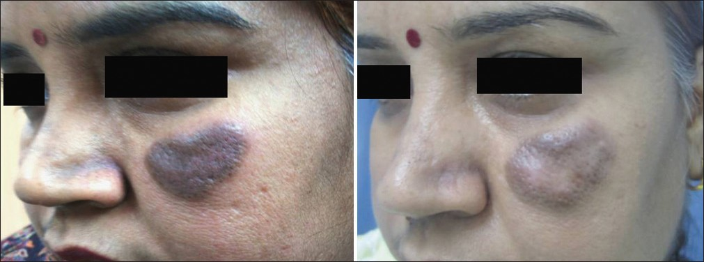
A 35-year-old female presented to our department with a 5 year history of single, asymptomatic, grey-brown pigmented, nodule over the left cheek [Figure 1]. It started as a pin head sized papule which gradually increased to 2.5 cm × 1.5 cm in size. Two years later similar lesions appeared on the forehead, both arms and upper back. There was no skin ulceration. No photosensitivity, fever or joint pain was present. Past and personal history was unremarkable.

- Before treatment – single, grey-brown nodule with prominent follicular orifices over left cheek. After treatment – residual lesion after three months of tacrolimus application
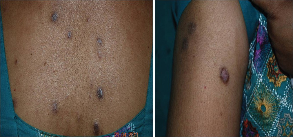
General physical and systemic examination was normal. Cutaneous examination revealed multiple, well-defined, grey-brown, indurated, non-tender plaques, varying in size from 0.5 cm × 0.5 cm to 1.5 cm × 2.5 cm, present on the left cheek, left forehead, both arms and upper back. Overlying surface showed prominent follicular openings, telangiectasia and peri-lesional erythema. Co-existent macular amyloidosis was present over upper back [Figure 2].

- Multiple grey-brown plaques over upper back
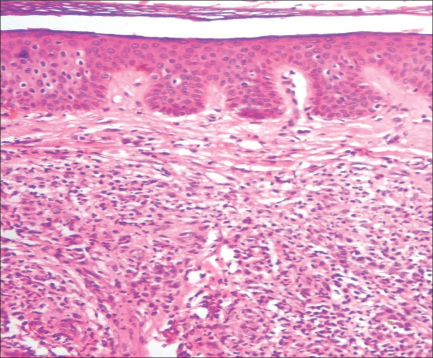
Routine hematological and biochemical investigations were normal. Skin biopsy (4 mm) from plaque revealed normal epidermis with clear sub epidermal ‘Grenz zone’ and pan dermal dense infiltrate comprising of neutrophils, lymphocytes, histiocytes and plasma cells. Small dermal vessels showed infiltration of neutrophils in the vessel wall along with peri-appendageal and peri-neural infiltrate in subcutaneous fat [Figure 3]. Features were consistent with diagnosis of GF.

- (H and E, 100 ×) Skin biopsy with normal epidermis and dense, mixed inflammatoey infiltrate beneath a narrow grenz zone in the dermis. Infiltrate is composed of mononuclear cells with neutrophils and eosinophils
She was started on intralesional triamcilone acetonide 10 mg/ml injection monthly with Tab. Dapsone 100 mg twice daily for about 1 year with no improvement. These were then stopped and cryotherapy was started. Six sessions of cryotherapy were performed once monthly after which she developed erythema and itching over the plaques and discontinued treatment. Topical tacrolimus 0.1% ointment twice daily was started. The lesions showed 40-50% improvement after 3 months of therapy [Figure 1]. The treatment was well tolerated without any side effects.
DISCUSSION
GF is an uncommon, benign, inflammatory dermatosis usually confined to the face. However, extrafacial lesions have also been reported. The aetiology is unknown.[1] Classically, red-brown or violaceous nodules or plaques with associated telangiectasia and follicular accentuation are seen on the face over sun-exposed sites. The condition is typically asymptomatic and has no systemic features.[1] The course is chronic and patient seek treatment due to cosmetic concerns.
Histology is usually diagnostic and should be performed to exclude other possible causes. Differential diagnosis includes lupus pernio, lupus vulgaris, lymphoma, discoid lupus erythematosus and deep mycotic infection. Skin biopsy is characterized by a mixed inflammatory infiltrate with a predominance of neutrophils and eosinophils in the dermis, in conjunction with small vessel vasculitis. There is a Grenz zone that separates the infiltrate from the epidermis and pilosebaceous units.[1]
The lesions are slow growing and tend to be persistent. The disease is notoriously resistant to therapies and often tends to relapse once the treatment is discontinued. Several medical and surgical modalities like topical and intralesional corticosteroids, cryotherapy, pulsed dye laser, PUVA, systemic corticosteroids, dapsone and antimalarials have been tried with variable success rates. Carbon dioxide laser has also been used in a case of recurrent GF.[2] Surgical excision has been performed with often unsatisfactory results.[3] Ablative procedures may leave residual pigmentation and scarring, whereas long-term application of corticosteroids is associated with skin atrophy, telangiectasia and other possible adverse effects.[3]
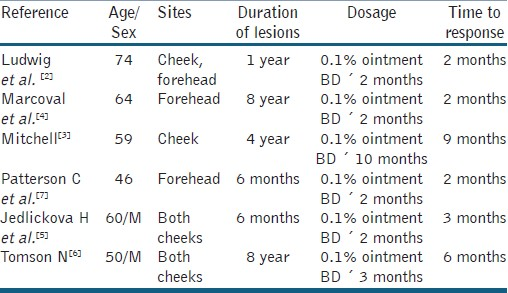
In recent years successes with topical calcineurin inhibitors has been reported. Several authors have reported complete or near-complete resolution of lesions after application of topical tacrolimus 0.1% ointment.[4–8] Treatment regimens, duration and time to resolution of lesions have varied in these case reports [Table 1]. The shortest time reported for resolution is 2 months after twice daily application. Others have found time to resolution to be between 4 and 6 months.[35] In our patient, treatment with tacrolimus 0.1% ointment twice daily for 3 months has resulted in improvement.

Tacrolimus inhibits T-cell proliferation, production and release of several pro-inflammatory cytokines like interleukin-2 (IL-2), IL-4, tumor necrosis factor-alpha, and interferon-gamma (IFN-gamma).[9] Although the pathogenesis is still unknown, it has been suggested that GF may be an IFN-gamma mediated disease.[10] In addition, an increased production of IL-5, probably induced by the clonal expansion of a locally recruited T-cell population may enhance the attraction of eosinophils into the lesions of GF.[11] Therefore, a possible mechanism of action of topical tacrolimus in this condition may be the inhibition of IFN-gamma and IL-5 production and release, induced by the down-regulation of the T-cell activity, primarily involving the calcineurin binding and inactivation. However, we did observe eosinophils in skin biopsy in our case, probably since the biopsy was taken after one year of oral dapsone.
Our patient experienced a relevant improvement within 3 months of treatment with tacrolimus ointment after no response with intra lesional steroids, dapsone and cryo therapy. In conclusion, the previous reports and our observation suggest that topical tacrolimus may be a well-tolerated, efficacious therapy for GF.
Source of Support: Nil
Conflict of Interest: None declared.
REFERENCES
- Granuloma faciale: A clinicopathologic study of 66 patients. J Am Acad Dermatol. 2005;53:1002-9.
- [Google Scholar]
- Recurrent Granuloma Faciale Successfully Treated with the Carbon Dioxide Laser. J Cutan Aesthet Surg. 2011;4:156-7.
- [Google Scholar]
- Successful treatment of granuloma faciale with tacrolimus. Dermatol Online J. 2004;10:23.
- [Google Scholar]
- Granuloma faciale: Treatment with topical tacrolimus. J Am Acad Dermatol. 2006;55:S110-1.
- [Google Scholar]
- Granuloma faciale successfully treated with topical tacrolimus: A case report. Acta Dermatovenerol Alp Panonica Adriat. 2008;17:34-6.
- [Google Scholar]
- Granuloma faciale treated successfully with topical tacrolimus. Clin Exp Dermatol. 2009;34:424-5.
- [Google Scholar]
- Granuloma faciale successfully treated with topical tacrolimus. Australas J Dermatol. 2009;50:217-9.
- [Google Scholar]
- Tacrolimus ointment: A review of its use in atopic dermatitis and its clinical potential in other inflammatory skin conditions. Drugs. 2005;65:827-58.
- [Google Scholar]
- Immunophenotypic analysis suggests that granuloma faciale is a c-interferon-mediated process. J Cutan Pathol. 1993;20:442-6.
- [Google Scholar]
- High local interleukin 5 production in granuloma faciale (eosinophilicum): Role of clonally expanded skin-specific CD4+ cells. Br J Dermatol. 2005;153:454-7.
- [Google Scholar]


