Translate this page into:
Giant Tuberous Xanthomas in a Case of Type IIA Hypercholesterolemia
Address for correspondence: Dr. Aniketh Venkataram, No 3437, 1st G cross, 7th main, Subbanna Gardens, Next to BTS garage, Vijaynagar, Bangalore - 560085, Karnataka, India. E-mail: anikethv@gmail.com
This is an open-access article distributed under the terms of the Creative Commons Attribution-Noncommercial-Share Alike 3.0 Unported, which permits unrestricted use, distribution, and reproduction in any medium, provided the original work is properly cited.
This article was originally published by Medknow Publications & Media Pvt Ltd and was migrated to Scientific Scholar after the change of Publisher.
Abstract
Tuberous xanthomas are papulonodular skin lesions present in lipoprotein metabolism disorders. A patient presented with multiple large swellings (up to 20 cm in size) all over the body, which on excision were found to be tuberous xanthomas. Investigations revealed the diagnosis of familial hypercholesterolemia. This case is reported to document the unusual presentation of giant tuberous xanthomas.
Keywords
Giant
hypercholesterolemia
lipoprotein
tuberous xanthomas
INTRODUCTION
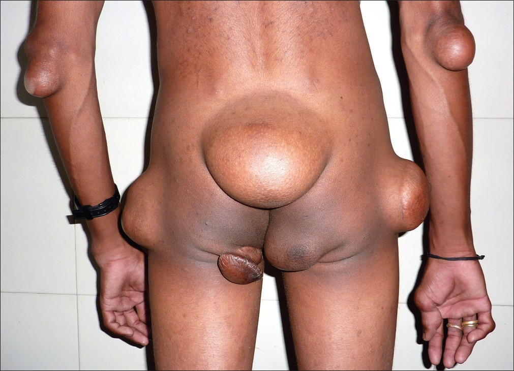
Xanthomas are benign plaques, papules, or nodules characterized by accumulation of lipid laden macrophages that develop in the cutis and subcutaneous tissue.[1] Tuberous xanthomas are firm painless yellow red nodules most commonly seen over extensor aspects of limbs and buttocks.[2] They are seen in several lipidoses and are usually indicative of a derangement in lipoprotein metabolism, in particular familial hypercholesterolemia and are usually not larger than 2 cm.[3] Recently a patient presented with multiple large swellings varying from 2 to 20 cm all over the body. On histopathological examination, these were revealed to be an atypical presentation of giant tuberous xanthomas, which is the first such reported case, to the best of our knowledge.
CASE REPORT
A 26-year-old nonconsanguineous male presented with multiple painless swellings over the body since 12 years with no significant family history. He first developed a swelling over the sacral region that progressed gradually to the present size of 20 cm × 15 cm × 10 cm [Figure 1]. At varying intervals, other swellings developed over his body at varying locations. The swellings were present over the buttocks, extensor aspects of elbows and knees and over the hands and feet and varied in size from 2 cm × 2 cm over dorsum of hands to 15 cm × 10 cm over buttocks. On examination, they were firm, nontender, mobile, and without any skin changes. Family history was unremarkable.

- Multiple swellings over extensor aspects of limbs and buttocks
Routine investigations including hemogram, X-rays were unremarkable. Fine needle aspiration cytology suggested spindle cell tumor. The sacral swelling was excised with overlying skin and primary closure was done. The gross specimen revealed a firm, homogenous grey white mass.
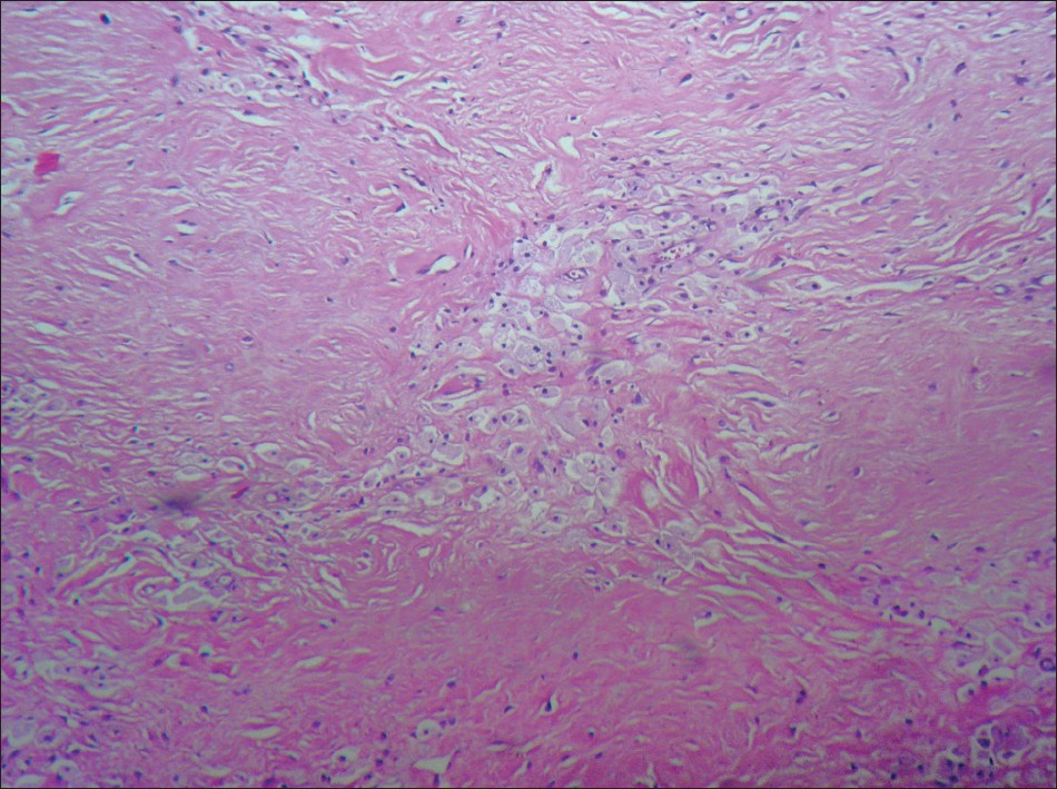
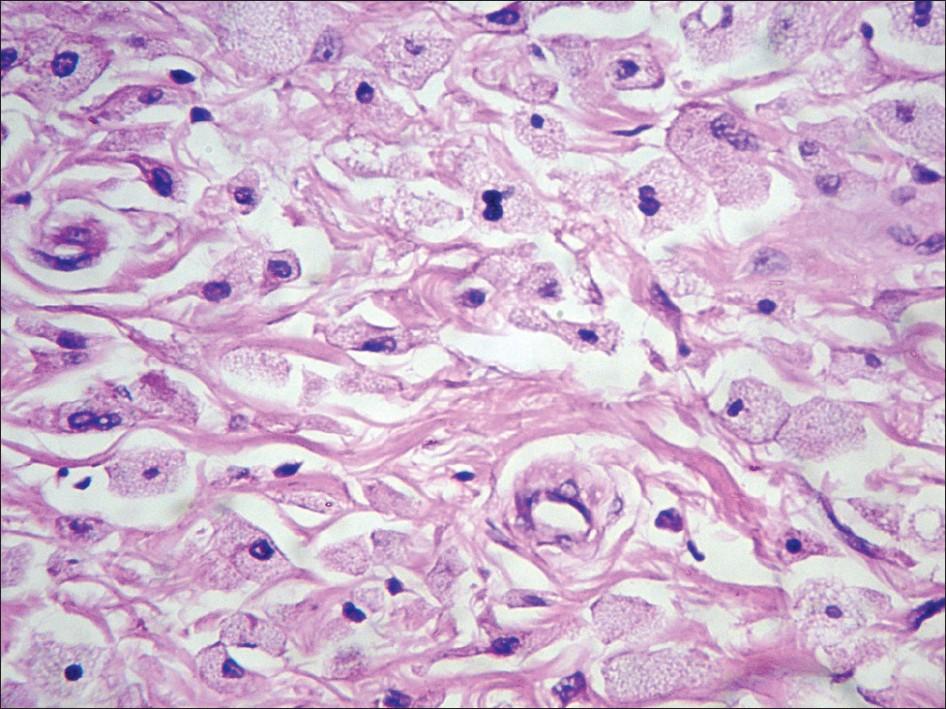
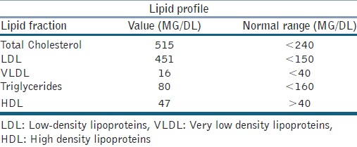
On histopathological examination, collections of foam cells and lipid laden macrophages with areas of fibrosis and cholesterol clefts were found indicative of tuberous xanthomas [Figures 2 and 3]. Lipid profile was performed that revealed raised levels of low-density lipoproteins (LDL) and total Cholesterol. Triglycerides and other lipoprotein fractions were normal, suggestive of type IIa familial hypercholesterolemia [Table 1].

- Histopathology showing foamy macrophages with fibrosis, cholesterol clefts

- Macrophages with intracellular lipid accumulation

The patient was started on lipid lowering medications including high dose atorvastatin with niacin. He was also worked up for other systemic manifestations of hypercholesterolemia including atherosclerosis and coronary artery disease. At present he is normolipaemic, and reports a generalized global reduction of the size of the xanthomas, with no development of any new lesions.
DISCUSSION
Xanthomas are lesions characterized by the accumulation of lipid laden macrophages. They occur when derangements in lipid metabolism lead to the leakage of lipids from the vasculature into the tissues, where they are phagocytosed by macrophages.[1] They have also been known to occur in normolipemic individuals.
Tuberous xanthomas are typically reddish-yellow papulonodular lesions of the skin. They are typically found over pressure areas such as extensor aspects of knees, elbows, and buttocks.[24] Histologically, apart from foamy macrophages, they have also been found to contain primitive mesenchymal cells, elongated perivascular, and fibroblast-like cells, and lysosome-filled macrophages, indicating possible stages in the evolution of dermal mesenchymal cells into mature, cholesterol-rich foam cells.[5]
Xanthomata are cutaneous markers of underlying lipid metabolism disorders, classified by Frederickson into five classes. Tuberous xanthomas are usually found in type IIa familial hypercholesterolemia.[36] Familial hypercholesterolemia is an autosomal codominant disorder with raised LDL levels due to increased production and reduced resorption of LDL secondary to dysfunctional LDL receptors. Heterozygotes express half the number of LDL receptors and homozygotes have between 0% and 25%.[7]
The consequences of such defects in LDL receptor genes are changes in vascular endothelial function, high serum total cholesterol and LDL cholesterol, with normal triglycerides. These are manifested as atherosclerosis and coronary artery disease.[78] The treatment options include changes in lifestyle and diet regulation, pharmacologic therapy and invasive procedures including lifelong lipid apheresis, and finally, liver transplantation.[9] Pharmacotherapy includes statins, bile acid sequestrants, ezetimibe, niacin that affect various steps in the pathways of cholesterol absorption and metabolism. The most common medication is a statin in combination with a cholesterol absorption inhibitor, and if needed a third drug such as a bile acid sequestrant.[10]
CONCLUSION
In conclusion, tuberous xanthomas are rarely larger than a few centimetres in size; however, in our patient they ranged up to 20 cm × 15 cm in size. This case was reported for the atypical presentation of multiple giant tuberous xanthomas which, to the best of our knowledge have not been reported thus far. They may be the only indicators of an underlying pernicious condition. Our experience demonstrates that they may present first to a surgeon, and a thorough workup is essential to identify the underlying condition in order to initiate early treatment to prevent later complications.
Source of Support: Nil.
Conflict of Interest: None declared.
REFERENCES
- Tuberous xanthomas in type IIA hyperlipoproteinemia. Indian J Dermatol Venereol Leprol. 2002;68:105-6.
- [Google Scholar]
- Dermal, subcutaneous, and tendon xanthomas: Diagnostic markers for specific lipoprotein disorders. J Am Acad Dermatol. 1988;19:95-111.
- [Google Scholar]
- Familial combined hypercholesterolemia type IIb presenting with tuberous xanthoma, tendinous xanthoma and pityriasis rubra pilaris-like lesions. Indian J Dermatol Venereol Leprol. 2010;76:293-6.
- [Google Scholar]
- Tuberous xanthoma in homozygous type II hyperlipoproteinemia. A histologic, histochemical, and electron microscopical study. Arch Pathol. 1975;99:293-300.
- [Google Scholar]
- New aspects of xanthomatosis and hyperlipoproteinemia. Curr Probl Dermatol. 1991;20:63-72.
- [Google Scholar]
- Familial hypercholesterolemia and coronary heart disease: A HuGE association review. Am J Epidemiol. 2004;160:421-9.
- [Google Scholar]
- Arterial mechanical changes in children with familial hypercholesterolemia. Arterioscler Thromb Vasc Biol. 2000;20:2070-5.
- [Google Scholar]
- Optimal management of familial hypercholesterolemia: Treatment and management strategies. Vasc Health Risk Manag. 2010;6:1079-88.
- [Google Scholar]
- A systematic review and meta-analysis of statin therapy in children with familial hypercholesterolemia. Arterioscler Thromb Vasc Biol. 2007;27:1803-10.
- [Google Scholar]


