Translate this page into:
Congenital Melanocytic Nevi: Catch Them Early!
Address for correspondence: Dr. Karthika Natarajan, Department of Dermatology, PSG Hospitals, Peelamedu, Coimbatore - 641 004, India. E-mail: karthikanatarajan@yahoo.com
This is an open-access article distributed under the terms of the Creative Commons Attribution-Noncommercial-Share Alike 3.0 Unported, which permits unrestricted use, distribution, and reproduction in any medium, provided the original work is properly cited.
This article was originally published by Medknow Publications & Media Pvt Ltd and was migrated to Scientific Scholar after the change of Publisher.
Abstract
We report a 2-week-old neonate with a large congenital melanocytic nevus over face treated with surgical curettage followed by a combination of carbon dioxide laser and Q-switched neodymium-doped yttrium aluminum garnet lasers. The results were satisfactory with near complete resolution after 1 year of age. This case is reported to emphasize the timely management and the importance of curettage prior to development of rete ridges.
Keywords
Congenital melanocytic nevus
curettage
neonate
INTRODUCTION
The congenital melanocytic nevus (CMN) is a type of melanocytic nevus found in infants atbirth and can occur in both sexes, with no known predilection. The CMN usually presents as a circumscribed, light brown to black patch or plaque, covering a surface area of any size involving any site of the body. The distribution of nevus over face is devastating and may carry co-morbidities in its long course. The nevi are classified according to the size of the lesion (in diameter): Small (<1.5 cm), medium (1.6-19.9 cm), and large (>20 cm). Many of these nevi may also present with hypertrichosis. The estimated prevalence for the largest forms of CMN is 0.002% of births.[1]
The predominant microscopic findings in CMN are collection of nevus cells upto deep dermis and subcutaneous fat including dermal appendages such as hair follicles, sebaceous glands, arrector pili muscles, and neurovascular structuresinclusive ofblood vessels walls. There is a relative absence of nevus cells in the subepidermal zone.[2–4]
We present a case of CMN which was treated with curettage and electrocautery in the initial session followed by a combination of Q-switched neodymium-doped yttrium aluminum garnet (Nd: YAG) and carbon dioxide (CO2) laser along with curettage.
CASE REPORT
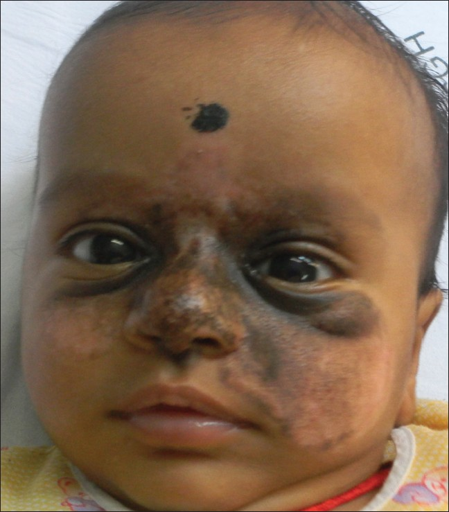
A 2-week-old male neonate of full-term normal vaginal delivery presented with CMN [Figure 1]. The nevus was present over the face involving lower forehead, the eyelids, nose, and both cheeks. On examination, the neonate was clinically normal. Under general anesthesia, we performed electrocautery and surgical curettage over the entire lesion except the eyelids in order to prevent trivial injuries to the eye. The wound was dressed with non-adherent antibiotic tulle (Sofra-tulle) and thisdressingwas removed after 48 h. The raw area showed crusting with ulceration which gradually healed with almost 60-70% clearance of the nevus [Figure 2].

- Fourteen-days-old neonate with congenital melanocytic nevus on face

- Three months infant following surgical curettage and dermabrasion
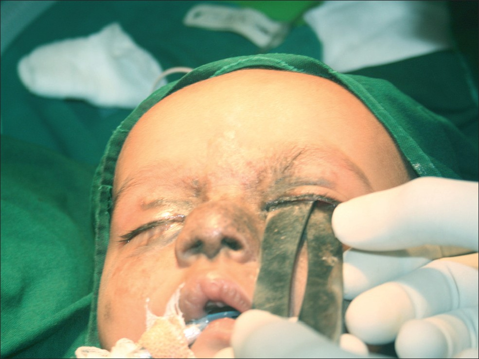
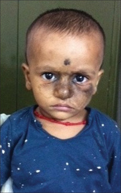
Six months later, the infant presented with residual lesion over the cheeks and nose and fine hairs over the cheek areas. The treatment was planned to performboth Q-switched Nd: YAG and CO2 lasers. Once again under general anesthesia with the help of an ophthalmologist to safeguard the eyeballs, a metallic flat shield was placed by lifting the upper eyelid since ophthalmic guard was not available [Figure 3]. The face of the infant was thoroughly wrapped with wet drapes during the procedure. The affected area was treated with Q-switched Nd: YAG laser (Medlite C6, Conbio Aesthetic Lasers, Freemont, USA) using the following settings: 1064 wavelength, 2J/cm2 fluence, 6 mm spot size, and repetition rate of 5. The settings of CO2 laser (Acupulse, Lumenis Ltd, Yokneam, Israel) under super pulse mode were fluence of 15J, spot size of 2 mm, and depth of 1 mm. During this treatment, the lesions over the eyelids weretreated with both lasers in addition to the residual lesions over cheek and nose. CO2 laser ablation was used by defocusing the beam to cause superficial damage. The post-operative period was uneventful with normal vision. Following this procedure, the lesion cleared upto 85-90% with minimal to no scarring [Figure 4].

- Protective metallic barrier as eye shield for laser surgery on table

- Outcome at 1 year of age
DISCUSSION
The management of CMN becomes mandatory for two reasons: One to reduce risk of malignant transformation especially in larger variants and second to enhance cosmesis.
Surgical excision of CMN depends on the size and site of the lesion. The larger lesions that require surgical excision are usually followed bytissue expanders, tissue grafts, and tissue flaps in order to repairthe large defects. Surgical curettage is relatively an alternate simpler procedure whenperformed during the neonatal period yields acceptable results.[5] The line of cleavage during neonatal stage is remarkably separable at the level of dermo-epidermal junction. The rationale of combining laser was to augment ablation using CO2 laser and Q-switched Nd: YAGlaser was to target both superficial and deep pigmentations.
Margulis et al.[6] successfully treated 44 patients of CMN involving the eyelids and periorbital region at the age of 6 months and above with excision and reconstruction. They also highlighted the significance of early intervention at neonatal period to avoid complicated procedures.
The lasers reported for CMN are high-energy pulsed CO2 laser, erbium: YAG laser, normal-mode ruby laser, Q-switched Nd: YAG laser, Q-switched ruby laser, and Q-switched alexandrite laser with varying outcomes.[7–11] The long-term recurrences with laser treatments cannot be excluded as a result oflack of penetration to deeper tissue levels.
The treatment of CMN, once established, becomes difficult and requires complicated surgical techniques. When the lesions are very large, such techniques cannot be used to remove the entire lesion.
Since the lesion is superficial during the neonatal stage, the lesions are more amenable to simpler surgical techniques such as electrocautery, CO2 laser ablation, and curettage. Early referral of such cases can lead to improved quality of life for the child.
Source of Support: Nil.
Conflict of Interest: None declared.
REFERENCES
- Congenital melanocytic nevi-when to worry and how to treat: Facts and controversies. Clin Dermatol. 2010;28:293-302.
- [Google Scholar]
- Congenital melanocytic nevi of the small and garment type. Clinical, histologic, and ultrastructural studies. Hum Pathol. 1973;4:395-418.
- [Google Scholar]
- A histologic comparison of congenital and acquired nevomelanocytic nevi. Arch Dermatol. 1985;121:1266-73.
- [Google Scholar]
- Curettage of giant congenital melanocytic nevi in neonates: A decade later. Arch Dermatol. 2002;138:943-7.
- [Google Scholar]
- Congenital melanocytic nevi of the eyelids and periorbital region. Plast Reconstr Surg. 2009;124:1273-83.
- [Google Scholar]
- Carbon dioxide laser dermabrasion for giant congenital melanocytic nevi. Plast Reconstr Surg. 2003;111:2209-14.
- [Google Scholar]
- Treatment of congenital nevomelanocytic nevi with the CO2 and Q-switched alexandrite lasers. Dermatol Surg. 2005;31:518-21.
- [Google Scholar]
- A neonate with a giant congenital naevus: New treatment option with the erbium: YAG laser. Br J Plast Surg. 2002;55:440-2.
- [Google Scholar]
- Effectiveness of the normal-mode ruby laser and the combined (normal-mode plus Q-switched) ruby laser in the treatment of congenital melanocytic nevi: A comparative study. Ann Plast Surg. 2002;49:476-85.
- [Google Scholar]


