Translate this page into:
A Study on Fractional Erbium Glass Laser Therapy Versus Chemical Peeling for the Treatment of Melasma in Female Patients
Address for correspondence: Dr. Neerja Puri, C/O Dr Asha Puri, House No 626, Phase Ii, Urban Estate, Dugri Road, Ludhiana, Punjab, India. E-mail: neerjaashu@rediffmail.com
This is an open-access article distributed under the terms of the Creative Commons Attribution-Noncommercial-Share Alike 3.0 Unported, which permits unrestricted use, distribution, and reproduction in any medium, provided the original work is properly cited.
This article was originally published by Medknow Publications & Media Pvt Ltd and was migrated to Scientific Scholar after the change of Publisher.
Abstract
Introduction:
Melasma is a commonly acquired hypermelanosis and a common dermatologic skin disease that occurs on sun-exposed areas of face.
Aims:
To assess the efficacy and safety of non-ablative 1,550 nm Erbium glass fractional laser therapy and compare results with those obtained with chemical peeling.
Materials and Methods:
We selected 30 patients of melasma aged between 20 years and 50 years for the study. The patients were divided into two groups of 15 patients each. Group I patients were subjected to four sessions of 1,550 nm Erbium glass non-ablative fractional laser at 3 weeks interval. In group II patients, four sessions of chemical peeling with 70% glycolic acid was performed.
Results:
After 12 weeks of treatment, percentage reduction in Melasma Area and Severity Index (MASI) score was seen in 62.9% in the laser group and 58.7% in the peels group.
Conclusion:
It was observed that 1,550 nm fractional laser is as effective as 70% glycolic acid peel in reducing MASI score in patients with melasma.
Keywords
Chemical peeling
erbium glass laser
glycolic acid
hyperpigmentation
laser
melasma
INTRODUCTION
Melasma is a common pigment disorder which causes significant emotional and psychosocial distress in patients.[1] It adversely affects the patients quality of life. Melasma is encountered in all skin types, but is particularly seen in women with Fitzpatrick skin types IV to VI. The pathogenesis of melasma is not fully understood. Genetic background and sun exposure seems to be the most important etiologic factors besides pregnancy, systemic drugs, hormonal medications and phototoxic or photoallergic cosmetics. Multiple etiologic factors have been implicated like high estrogen states (pregnancy, oral contraceptives), cosmetics and autoimmune thyroid disease. Sunlight exposure appears to be essential for its development. Melasma is often difficult to manage because of its refractory and recurrent nature.[2] The results of laser therapy and intense pulsed light therapy in melasma are generally disappointing and treatment is limited by adverse affects such as the occurrence of post inflammatory hyperpigmentation, especially in dark-skinned patients. Therefore, the use of these devices is controversial. Recently, non-ablative fractional laser therapy at 1,550 nm was reported as a treatment for melasma.[34] At this wavelength, water absorption is predominant. Chemical peels are used to create an injury of a specific skin depth with the goal of stimulating new skin growth and improving surface texture and appearance.[5] The exfoliative effect of chemical peels stimulates new epidermal growth and collagen with more evenly distributed melanin.
Melasma is a symmetric progressive hyperpigmentation of the facial skin that occurs in all races but has a predilection for darker skin phenotypes.[678] Clinically, melasma can be divided into centrofacial, malar and mandibular, according to the pigment distribution on the skin. By Wood's light examination, melasma can be classified into epidermal, dermal or mixed type. Chemical peeling has a low rate of complications and is popular due to the low costs involved and to a technique which is easy to learn.[91011]
In fractional laser therapy, multiple small coagulated zones are separated by surrounding untreated tissue.[1213] It was reported that these microscopic treatment zones allow transport and extrusion of microscopic epidermal necrotic debris including melanin from melanocytes through a compromised dermoepidermal junction.[1415] Generally, a visible wound does not appear because these microscopic treatment zones have a diameter less than 100 μm. The stratum corneum was found to be intact after 24 hours. The recovery is relatively fast because only part of the skin surface is treated in one session.[16]
Aims
To assess the efficacy and safety of non ablative 1,550 nm fractional laser therapy and compare results with those obtained with chemical peeling.
MATERIALS AND METHODS
We selected 30 patients of melasma aged between 20 years and 50 years for the study. The patients were divided into two groups of 15 patients each. Group I patients were subjected to four sessions of 1,550 nm Erbium glass fractional laser at 3 weeks interval. In group II patients, four sessions of chemical peeling with 70% glycolic acid was performed. The patients were randomised on the basis of willingness to undergo laser treatment and the non laser group was informed about the laser treatment option also available. The peels were performed every 3 weekly for a maximum of four sessions. Written informed consent from all the patients was taken before the start of the study and risks, benefits and potential complications were communicated to the patients. Prior approval of hospital ethical committee was taken for the study. The study received ethical committee clearance. All the participants were subjected to Wood's light to determine the type of melasma (epidermal from dermal or mixed. Melasma Area and Severity Index (MASI) score was calculated in all the patients at the beginning of each session. All patients were instructed to use sunscreen (sun protection factor between 30 and 50) every 3 hours when outside. Improvement was also assessed by serial photographs as assessed by the physician. A 1,550 nm Erbium glass non-ablative laser was used. Anaesthesia for laser therapy consisted of topical 2.5% lidocaine and 2.5% prilocaine ointment applied an hour before each treatment. Each treatment session involved eight fractional laser passes to create an estimated final density of ~ 2,000-2,500 macroscopic treatment zones/cm2. Four passes were made in one direction and four perpendicularly. The energy per micro beam was 10 mj. Skin type II and III were treated at ~ 20% coverage and skin type IV and V at ~ 14% coverage. The follow-up visits were scheduled at 3 weeks, 3 months and 6 months after the last treatment day.
The patients on chemical peeling were strictly instructed to apply sun block cream, during and after therapy along with emollients in unlimited quantities. All side effects were documented and patients were asked to score erythema, oedema, crusting and blistering on a scale from zero to three. Patients were asked to score the improvement of hyperpigmentation on a visual analog scale from zero to 10, with zero as no improvement and 10 as the best possible improvement (Patients Global Assessment [PGA]). Patients were also asked whether they would recommend the treatment to their friends and colleagues. Pain in the laser group was recorded on a scale from zero to 10 (visual analog scale) after the first and the third treatment session.
The following patients were included in our study:
-
All patients between age group of 20-50 years
-
Patients with epidermal and dermal type of melasma
-
Patients with Fitzpatrick skin types II to V having moderate to severe melasma
-
Patients with mental capacity to give informed consent.
The following patients were excluded from our study:
-
Participants with a history of hypertrophic scars or keloids
-
Participants with recurrent herpes infection
-
Presence of active dermatitis
-
Patients with unrealistic expectations
-
Patients with use of isotretinoin in past 6 months
-
Patients with history of prior treatment.
RESULTS
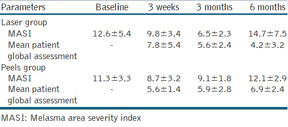
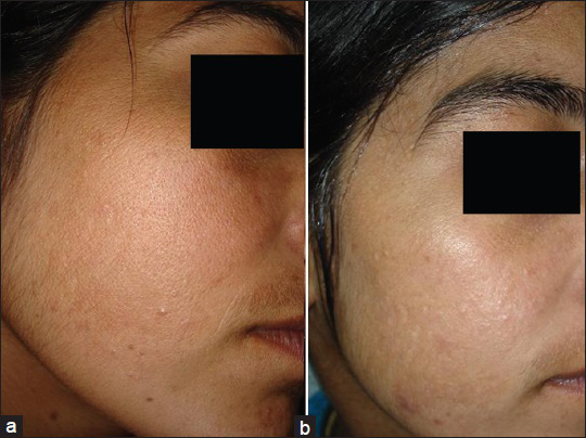
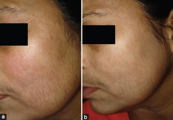
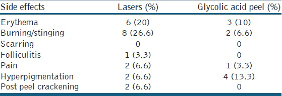
Table 1 shows that the average decrease in MASI score in both the groups was found to be statistically highly significant. However, the comparison of decrease in MASI score between both the groups was not statistically significant (P > 0.005) Regarding the age distribution of patients, it was seen that maximum (60%) patients were between 31 years and 40 years, 26.6% patients were between 41 years and 50 years and 13.35% patients were between 20 years and 30 years of age. Females outnumbered males and female: male ratio was 6.5:1. The pattern of melanosis was of malar type in 40% patients, mixed in 40% patients and centrofacial in 20% patients. It was seen that epidermal type of melasma was seen in 70% patients, dermal was seen in 10% patients and mixed melasma was seen in 10% patients. Regarding the skin types, 43.3% patients had skin type IV, 36.6% patients had skin type III and 20% patients had skin type II. It was seen that the commonest cause of melasma in our study was after pregnancy in 40% patients, oral contraceptive use was associated with melasma in 30% patients and outdoor occupation was the cause of melasma in 30% patients. Subjective response as graded by the patient showed good or very good response in 70% patients in laser group [Figures 1a and b] and 64% in the glycolic acid group [Figures 2a and b], which was statistically insignificant. The commonest side effect after peels [Table 2] was burning which was seen in 26.6% patients in laser group, where as in glycolic acid peel burning sensation was seen only in 6.6% patients. Post peel erythema was seen in 20% patients with lasers and 10% patients with glycolic acid peel. Pain as a side effect was seen in 6.6% patients with lasers and 3.3% patients with glycolic acid peel. Hyperpigmentation was seen in 13.35% patients with laser group and 6.6% patients with glycolic acid peels. Post peel crackening was seen in 6.6% patients with lasers and none of the patients with glycolic acid peel. After treatment patients were asked to evaluate the discomfort from the two different peeling solutions. They found the lasers caused more discomfort and slight pain, whereas 70% glycolic acid peel caused strong stinging during the application, excessive desquamation during the next 4-5 days, which interfered with patients daily activities. The glycolic acid procedure was associated with stinging and burning, which were most pronounced during the first procedure.


- (a and b) Pre and post treatment photograph of a 22-years-old patient before and after laser therapy

- (a and b) Pre and post treatment photograph of a 40-years-old patient before and after peeling with 70% glycolic acid

DISCUSSION
After 12 weeks of treatment, percentage reduction in MASI score was seen in 62.9% in the laser group and 58.7% in the peels group. Physician Global Assessment (PhGA) improved (P < 0.001) in both groups at 3 weeks. Mean treatment satisfaction and recommendation were significantly higher in the laser group at 3 weeks (P < 0.05). However, melasma recurred in both the groups after 6 months. In both groups, the PGA showed a distinct improvement at the 3-week follow-up (P < 0.001). The PGA and MASI showed no statistically significant differences within or between the groups. Clinically, recurrence of melasma was encountered in the majority of both patient groups at the 6-month follow-up. Non-ablative 1,550 nm fractional laser therapy proved to be a safe treatment option for patients including those with darker skin types (Fitzpatrick skin type IV and V). The patients considered non-ablative 1,550 nm fractional laser therapy to be a satisfactory and recommendable treatment.
There are many treatment modalities for melasma; however, there is no sure-fire method of treating this disease. Chemical peels remain popular for the treatment of some skin disorders and for aesthetic improvement. Patients who are willing to undergo continued treatment are likely to be the best candidates. Clinicians should remember that there can be excellent synergy between peels and other procedures. Finally, it is important for patients to maintain a good sun protection regimen to optimize the clinical results achieved with chemical peels. The main limitations of our study are: (1) A small number of included patients; (2) a sample size that was powered for PhGA only; (3) laser settings that may have been sub optional; and (4) a possible difference in the motivation and the therapy adherence between the two groups.
CONCLUSIONS
Initially, good results were seen at the short-term follow-up. However, in both groups pigmentation worsened during the course of time. No significant differences were found between the two groups on longer follow-up and the results at 6 months. In conclusion, in this study, non-ablative 1,550 nm fractional laser therapy proved to be a safe treatment option for patients with melasma, including those with darker skin types. Patients considered laser therapy to be satisfactory and recommendable.
Source of Support: Nil
Conflict of Interest: None declared.
REFERENCES
- Melasma and its impact on health-related quality of life in Hispanic women. J Dermatol Treat. 2007;18:5-9.
- [Google Scholar]
- Treatment of melasma: A review of clinical trials. J Am Acad Dermatol. 2006;55:1048-65.
- [Google Scholar]
- Utilizing fractional resurfacing in the treatment of therapy-resistant melasma. J Cosmet Laser Ther. 2005;7:39-43.
- [Google Scholar]
- Glycolic acid peeling in the treatment of acne. J Eur Acad Dermatol Venereol. 1999;12:119-22.
- [Google Scholar]
- The treatment of melasma with fractional photothermolysis: A pilot study. Dermatol Surg. 2005;31:1645-50.
- [Google Scholar]
- Fractional photothermolysis: A new concept for cutaneous remolding using microscopic patterns of thermal injury. Lasers Surg Med. 2004;34:426-38.
- [Google Scholar]
- Laser-induced transepidermal elimination of dermal content by fractional photothermolysis. J Biomed Opt. 2006;11:41-8.
- [Google Scholar]
- The prevalence and risk factors of post inflammatory hyperpigmentation after fractional resurfacing in Asians. Lasers Surg Med. 2007;39:381-5.
- [Google Scholar]


