Translate this page into:
Pedunculated superficial angiomyxoma of the thigh mimicking molluscum pendulum
*Corresponding author: Noureddine Litaiem, Department of Dermatology, Charles Nicolle Hospital, Tunis 1006, Tunisia. noureddine.litaiem@gmail.com
-
Received: ,
Accepted: ,
How to cite this article: Hawilo A, Fakhfakh W, Litaiem N. Pedunculated superficial angiomyxoma of the thigh mimicking molluscum pendulum. J Cutan Aesthet Surg. doi: 10.4103/JCAS.JCAS_72_23
Abstract
Superficial angiomyxoma (SAM) is a rare acquired benign neoplasm. It may be sporadic or associated with Carney’s complex. Typical locations for SAM are the head, neck, and trunk. It usually presents as a skin-colored papule or sessile lobulated nodule. Large pedunculated forms are less common. We describe a sporadic massive pedunculated SAM of the thigh occurring in a 75-year-old man and provide a comprehensive summary of SAM characteristics.
Keywords
Acrochordon
Molluscum pendulum
Myxoma
Superficial angiomyxoma
INTRODUCTION
Superficial angiomyxoma (SAM), also known as cutaneous myxoma, is a rare acquired benign neoplasm. It may be sporadic or arise in association with syndromes such as Carney complex.1
SAM is usually located on the trunk, head, and neck. Genital and acral locations have been described. It usually appears as a sessile nodule. We report herein an unusual case of pedunculated SAM of the thigh and provide a comprehensive summary of its characteristics.
CASE REPORT
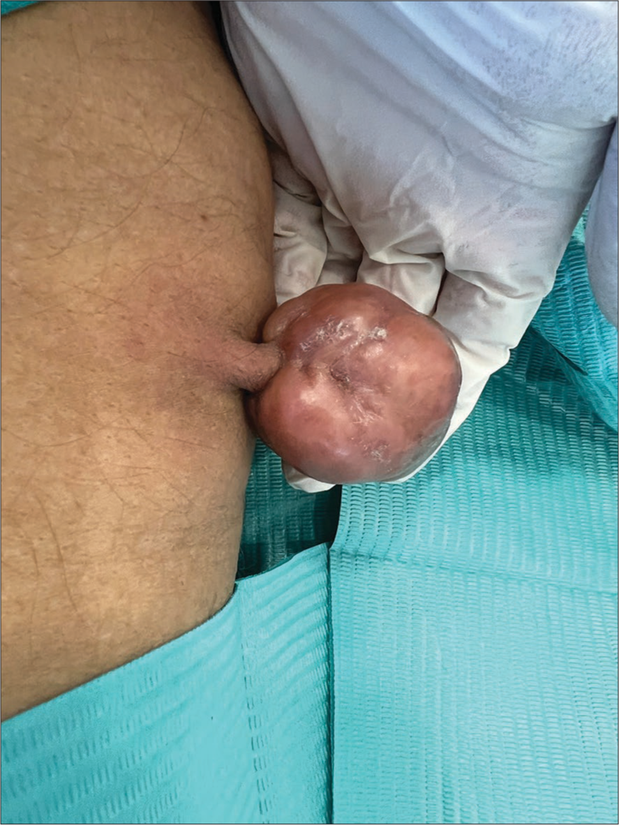
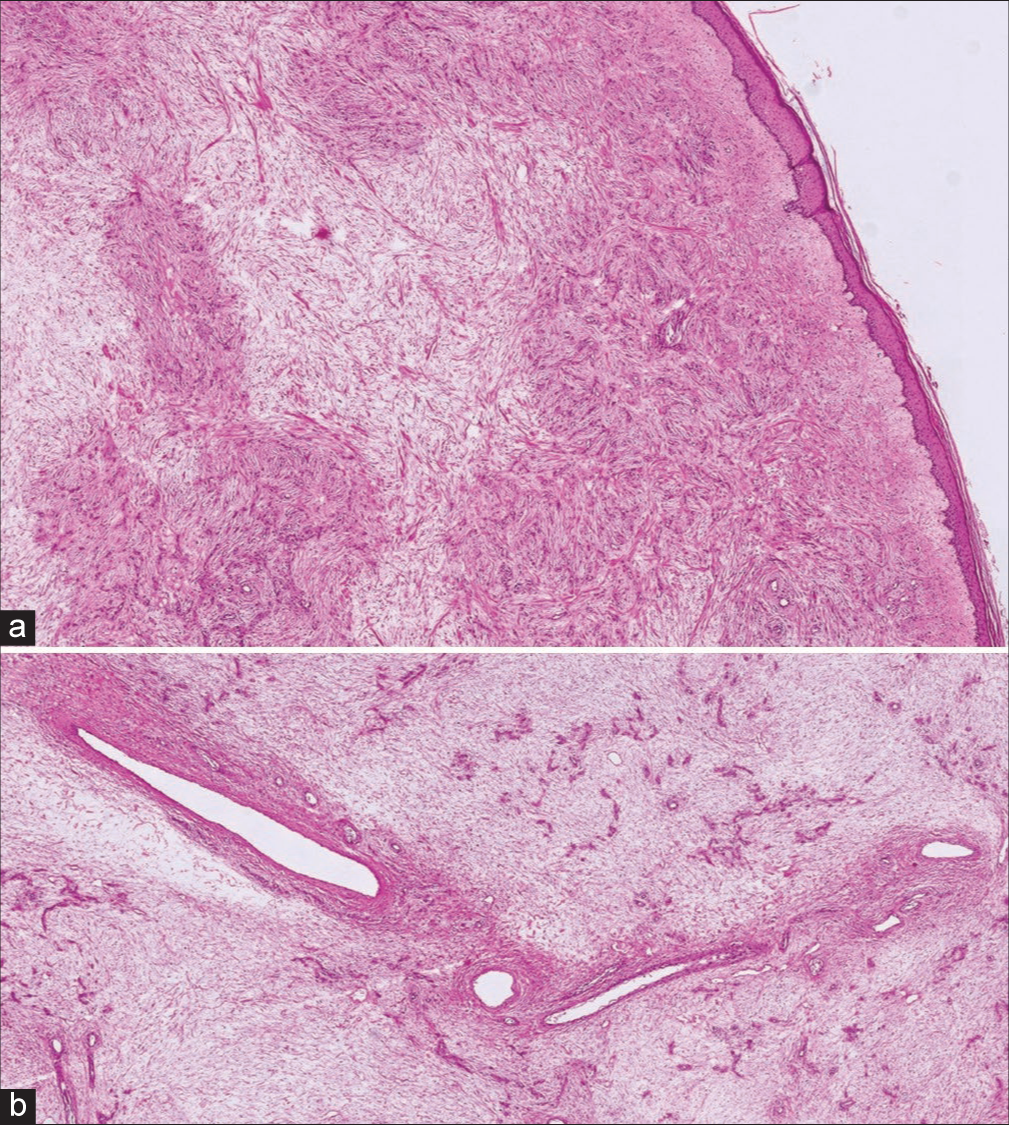
A 75-year-old man with a medical history of diabetes mellitus presented with a 3-year history of a slow-growing asymptomatic tumor of the inner aspect of the right thigh. Clinical examination showed a 6 cm × 5 cm × 3 cm firm, skin-colored, well-circumscribed, pedunculated tumor with a smooth surface with no associated lymphadenopathy [Figure 1]. Physical examination was otherwise unremarkable. A shave excision of the tumor was performed. No unusual bleeding was encountered during surgery and no clamping at the base of the tumor was performed before excision. Histopathological examination showed a polypoid superficial paucicellular tumor with spindle cells in a myxoid loose matrix associated with blood vessels of variable size [Figure 2]. There was no evidence of cellular atypia or malignancy. The diagnosis of SAM was established. Thoraco- abdomino-pelvic computed tomography scan and bone scintigraphy were normal. No recurrence was noted after 6 months of follow up.

- Clinical aspect: A 6 cm × cm 5 × 3 cm firm, skin-colored, well-circumscribed, and pedunculated tumor on the right thigh.

- Histopathological examination (a) H&E ×40, (b) H&E ×80. Polypoid superficial paucicellular tumor with spindle cells in a myxoid loose matrix associated with blood vessels of variable size. H&E: Hematoxylin and eosin.
DISCUSSION
In 1998 Allen et al.2 reported several cases of SAM without evidence of Carney’s complex. SAM is a rare slow-growing cutaneous neoplasm that usually measures 1–5 cm, but larger variants have been recorded.2 It can appear at any age, with a peak of incidence in the fourth and fifth decades of life.
Given its location, SAM in our patient can mimic other tumors. In our patient, the aspect of a skin-colored pedunculated tumor of the thigh can be confused with a giant molluscum pendulum/acrochordon. SAM should be distinguished from aggressive angiomyxoma. The latter tends to grow deeper into the tissue and carries a high risk of recurrence after surgery.3 The other differential diagnoses in men include acrotesticular neoplasms, inguinal hernia, hydrocele, angiomyofibroblastoma, and myxoid sarcoma.
The diagnosis of SAM is based on histopathological examination that shows lobulated, well-defined markedly hypocellular mesenchymal proliferation characterized by a mucinous matrix within the dermis and the subcutis, with spindled to stellate cells and thin vessels of variable size.1 Mitoses and nuclear atypia are usually absent. The immunohistochemical staining is generally positive for CD34, S100 protein, SMA, muscle-specific actin, and factor XIIIa, while pancytokeratin, desmin, and MUC4 stains are typically negative.4
Surgical excision is the treatment of choice. Local recurrence may occur after surgery, but not metastasis. Recurrence rates are variable, ranging between 3.6% and 38.1%, occurring when excision is incomplete.1,2,5 Mohs micrographic surgery could be proposed for difficult cases.5 Complete excision is required to avoid recurrences but no recommended minimal surgical margins are defined. Nonetheless, in the largest clinicopathological series of SAM, shave excision was performed in 32/54 cases with no recurrences.1 In our case, given the pedunculated aspect of the tumor, shave excision was indicated with no recurrence at 6 months. A long-term follow-up is necessary because recurrences can appear years after surgery.
CONCLUSION
Superficial angiomyxoma is a rare and benign myxoid tumor usually appearing as a 1–5 cm sessile nodule. This case illustrates a rare tumor in men occurring with an unusual presentation. Rare clinical presentations including giant and pedunculated SAM should not be misdiagnosed. This differential should be considered mainly in the differential diagnosis of pelvic and perineal masses. Shave excision is a good treatment option for pedunculated SAM that may lead to good cosmetic outcome and low recurrence rate.
Authors’ Contributions
All authors had access to the data and a role in writing this manuscript.
Declaration of patient consent
The authors certify that they have obtained all appropriate patient consent forms. In the form, the patient(s) has/have given his/her/their consent for his/her/their images and other clinical information to be reported in the journal. The patients understand that their names and initials will not be published and due efforts will be made to conceal their identity, but anonymity cannot be guaranteed.
Conflict of interest
There are no conflicts of interest.
Financial support and sponsorship
Nil.
References
- A clinicopathologic analysis of 54 cases of cutaneous myxoma. Hum Pathol. 2022;120:71-6.
- [CrossRef] [PubMed] [Google Scholar]
- Superficial angiomyxomas with and without epithelial components: Report of 30 tumors in 28 patients. Am J Surg Pathol. 1988;12:519-30.
- [CrossRef] [PubMed] [Google Scholar]
- Aggressive angiomayxoma in men: Case report and systematic review. Ann Med Surg (Lond). 2022;79:103880.
- [CrossRef] [PubMed] [Google Scholar]
- A rare case of vulvar superficial angiomyxoma in a pediatric patient. J Pediatr Adolesc Gynecol. 2020;33:727-9.
- [CrossRef] [PubMed] [Google Scholar]
- Mohs micrographic surgery for the treatment of superficial angiomyxoma. Dermatol Surg. 2016;42:1014-6.
- [CrossRef] [PubMed] [Google Scholar]


