Translate this page into:
Purpura during dermabrasion: A false endpoint in vitiligo surgery over atrophic lesions
*Corresponding author: Suman Patra, Department of Dermatology Venereology and Leprology, All India Institute of Medical Sciences (AIIMS) Jodhpur, Jodhpur, Rajasthan, India. patrohere@gmail.com
-
Received: ,
Accepted: ,
How to cite this article: Patra S, Kumar S, Kaur M, Mandiya S. Purpura during dermabrasion: A false endpoint in vitiligo surgery over atrophic lesions. J Cutan Aesthet Surg. 2025;18:66-7. doi: 10.4103/JCAS.JCAS_215_22
Dear Editor,
Preparation of the recipient area remained one of the crucial steps in the surgical management of vitiligo. Among the various methods of recipient area preparation, dermabrasion with either manual or motorized dermabrader is the standard procedure. The endpoint of dermabrasion is defined as the appearance of pinpoint bleeding spots, which roughly corresponds to the level of the papillary dermis.
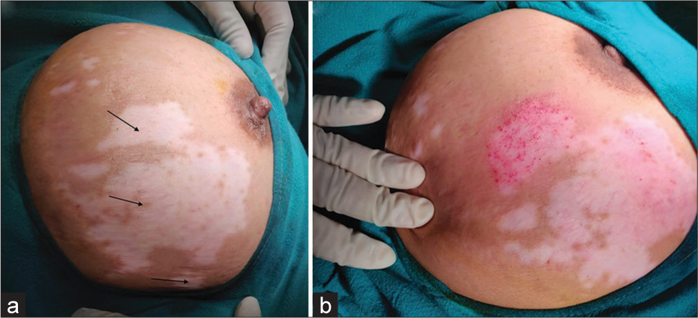
While doing dermabrasion in a stable vitiligo lesion over the breast [Figure 1a] in a young woman before noncultured epidermal suspension transfer technique, we observed an interesting finding. The skin of that area was atrophic along with the presence of multiple striae due to the prolonged application of topical corticosteroid preparations over the counter. While dermabrading the area with a manual dermabrader, after the initial attempt there was the appearance of many purpuric macules [Figure 1b]. The tiny red dots mimic exactly those of pinpoint bleeding spots as the endpoint of dermabrasion. The lesions appeared very quickly after initial attempt of dermabrasion and were not blanchable. They could not be rubbed off from the surface [Video S1] as expected in pinpoint bleeding of dermabrasion. We noticed a similar occurrence during the dermabrasion of striae in another patient earlier too. There was no prior history of bleeding diathesis in the patient. The complete blood count including platelet count, prothrombin time, and activated partial thromboplastin time was within normal limits. There is no report of a similar occurrence in the literature. The fragile vessels at the dermis beneath the atrophic epidermis lacks connective tissue support. Mechanical dermabrasion may lead to mechanical damage to the capillaries leading to rupture of the wall and causing purpura. In such circumstances, other methods of recipient area preparation like using chemically assisted gauze dermabrasion,1 cryotherapy, radiofrequency ablation, and carbon dioxide laser might be useful.2 To identify the adequate depth of dermabrasion we can take the surrounding normal skin adjacent to the atrophic lesion as a comparator. The depth of the dermabrasion should be uniform and merging with the dermabraded adjacent skin.

- (a) Vitiligo lesion over the right breast with multiple striae (black arrow); (b) purpuric macules following attempt of dermabrasion with manual dermabrader.
Authors’ contributions
All the authors; Dr. Suman Patra, Dr. Shubham Kumar, Dr. Maninder Kaur and Dr. Shubham Mandiya, have contributed to the research study. The concepts, design, definition of intellectual content, literature search, clinical studies, manuscript preparation, manuscript editing and manuscript review are carried by all the authors. Dr. Suman Patra has contributed to data acquisition while Dr. Shubham Mandiya has taken the role of guarantor.
Ethical approval
Institutional Review Board approval is not required.
Declaration of patient consent
The authors certify that they have obtained all appropriate patient consent.
Conflicts of interest
There are no conflicts of interest.
Use of artificial intelligence (AI)-assisted technology for manuscript preparation
The authors confirm that there was no use of artificial intelligence (AI)-assisted technology for assisting in the writing or editing of the manuscript and no images were manipulated using AI.
Financial support and sponsorship: Nil.
References
- Chemically assisted gauze abrasion: An economical and convenient alternative to mechanical dermabrasion in vitiligo surgery. J Am Acad Dermatol 2021:S0190-9622(21)02185-X.
- [CrossRef] [Google Scholar]
- Role of recipient-site preparation techniques and post-operative wound dressing in the surgical management of vitiligo. J Cutan Aesthet Surg. 2015;8:79-87.
- [CrossRef] [PubMed] [Google Scholar]


