Translate this page into:
The Rhombic Bilobed Flap, a Simple, Geometrically Designed Flap
Address for correspondence: Yoshiaki Sakamoto, Department of Plastic and Reconstructive Surgery, Keio University School of Medicine, 35 Shinanomachi, Shinjuku-ward, Tokyo - 160-8582, Japan. E-mail: ysakamoto@z8.keio.jp
This is an open-access article distributed under the terms of the Creative Commons Attribution-Noncommercial-Share Alike 3.0 Unported, which permits unrestricted use, distribution, and reproduction in any medium, provided the original work is properly cited.
This article was originally published by Medknow Publications & Media Pvt Ltd and was migrated to Scientific Scholar after the change of Publisher.
Abstract
We describe a combination of the common rhomboid flap and bilobed flap and provide an example of its use. The rhombic bilobed flap is simple to use and is associated with fewer complications, such as pin-cushioning and standing cone deformities, while minimizing the risk of skin necrosis and tension on the flap.
Keywords
Bilobed flap
rhomboid flap
skin cancer
skin flap
INTRODUCTION
The goals of facial reconstruction surgery centre on closing defects in an inconspicuous manner. Fortunately, the robust vascular supply of the face allows for many reconstructive options using localized flaps. The rhomboid flap is commonly used for facial skin defects because of its simple design. However, a severe transposition may result in the development of a standing cone deformity, making closure of the flap difficult as a result of the tension on the tissue. If the raised flap region is closed, severe tension will also lead to the development of other deformities that affect facial symmetry. To solve this problem, we have employed a modification of the bilobed flap.
TECHNIQUE
To create the modified rhombic bilobed flap design, an ABCD rhombus, with angles of 60 and 120 degrees, is drawn around the defect. The first lobe of the bilobed flap is formed by constructing a standard Dufourmentel type of rhombic flap[1] and the BAD angle is the same as the DEF angle, which makes the first lobe of the bilobed flap equal to the size of the defect. The second lobe of the flap is created from half to two-thirds of the first lobe. The angle of FGH is also equal to the angles of BAC [Figure 1].

- Design of the bilobed rhombic flap
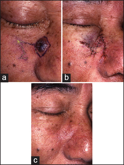
A representative case of a 68-year-old man with a 9 mm, benign nevus on the side of his nose is described. One-year post-operatively, the ectropion of the inferior eyelid and the nasal deformities were no longer apparent; the patient was satisfied with the result [Figure 2].

- Defect reconstruction. (a) Nevus cell nevus on the side of the nose and the planning of the flap (b) immediately after the operation And the (c) 1-year post-operative result
DISCUSSION
The original rhombic bilobed flap concept required that the lengths of the sides of the flaps be the same but that the angles at the tips of the flaps be small.[23] When using this approach, necrosis was noted at the tips of the second lobe because of its greater distance from the base of the flap. Therefore, we changed the design so that the angles at the vertices of the flaps were the same. This allowed the sides of the first lobe, around the defect, to be the same but the lengths of the sides of the second lobe to be shorter. Using this design, we did not note any skin necrosis.
The bilobed flap design has advantages over the single flap design in that the tension and stress on the skin can be dispersed across two flaps rather than across the single flap.[4] A standard bilobed flap has the disadvantage of pin-cushioning that often develops as a result of the curvilinear flaps; the incidence of pin-cushioning is reported to be 5%.[5] The precise geometric design of the revised rhombic bilobed flap decreases the incidence of pin-cushioning by producing angular corners.
CONCLUSION
In conclusion, the revised rhombic bilobed flap is very simple to use and is associated with fewer complications, including pin-cushioning and standing cone deformities, while minimizing the risk of skin necrosis and tension on the flap.
Source of Support: None of the authors has a financial interest in any of the products, devices or drugs mentioned in this manuscript.
Conflict of Interest: The authors report no conflicts of interest.
REFERENCES
- Modern trends in plastic surgery. Design of local flaps. Mod Trends Plast Surg. 1966;2:38-61.
- [Google Scholar]
- Stalked local rhinoplasty with bilobed flap, cover the secondary defect from the first lobe by the secondary lobe. German for surgery. 1918;143:385.
- [Google Scholar]
- Reconstruction of the nose utilizing a bilobed flap. Int J Dermatol. 1994;33:657-60.
- [Google Scholar]


