Translate this page into:
Angiomatosis: A Rare Vascular Proliferation of Head and Neck Region
Address for correspondence: Prof. Sujata Jetley, Department of Pathology, Hamdard Institute of Medical Sciences and Research, Jamia Hamdard, New Delhi - 110 062, India. E-mail: drjetley2013@gmail.com
This is an open-access article distributed under the terms of the Creative Commons Attribution-Noncommercial-Share Alike 3.0 Unported, which permits unrestricted use, distribution, and reproduction in any medium, provided the original work is properly cited.
This article was originally published by Medknow Publications & Media Pvt Ltd and was migrated to Scientific Scholar after the change of Publisher.
Abstract
Angiomatosis is a diffuse vascular lesion which clinically mimics hemangioma or vascular malformation. It usually involves multiple tissues and is histopathologically characterised by proliferation of vessels of varying calibre intimately admixed with large amount of adipose tissue. Its surgical removal is very difficult because of its infiltrative nature. Therefore, a precise histopathological diagnosis is of utmost importance. It is usually seen in females in the first two decades and commonly involves lower extremities. Angiomatosis of head and neck region is very rare. Here we present a rare case of angiomatosis of the lower face involving right cheek and lip in a 4-year-old boy clinically diagnosed as hemangioma. Histopathological differential diagnosis of angiomatosis is also discussed.
Keywords
Angiomatosis
head
neck
hemangioma
histopathological diagnosis
INTRODUCTION
Angiomatosis is a rare vascular lesion characterised by diffuse proliferation of blood vessels with accompanying mature adipose tissue. It affects a large segment of the body in a contiguous fashion either by vertically involving multiple tissue types (e.g., subcutis, muscle, bone) or by involving similar tissue types (e.g., multiple muscles).[1] It is primarily seen in the first two decades of life with a slight female predilection. Surgical removal of angiomatosis is very difficult and is associated with a high recurrence rate.
Angiomatosis is usually seen in lower extremities followed by the chest wall, abdomen and upper extremity. Very few cases are reported from the head and neck region.[2] We report an unusual case of angiomatosis seen in the face of a 4-year-old boy with an initial clinical diagnosis of hemangioma.
CASE REPORT
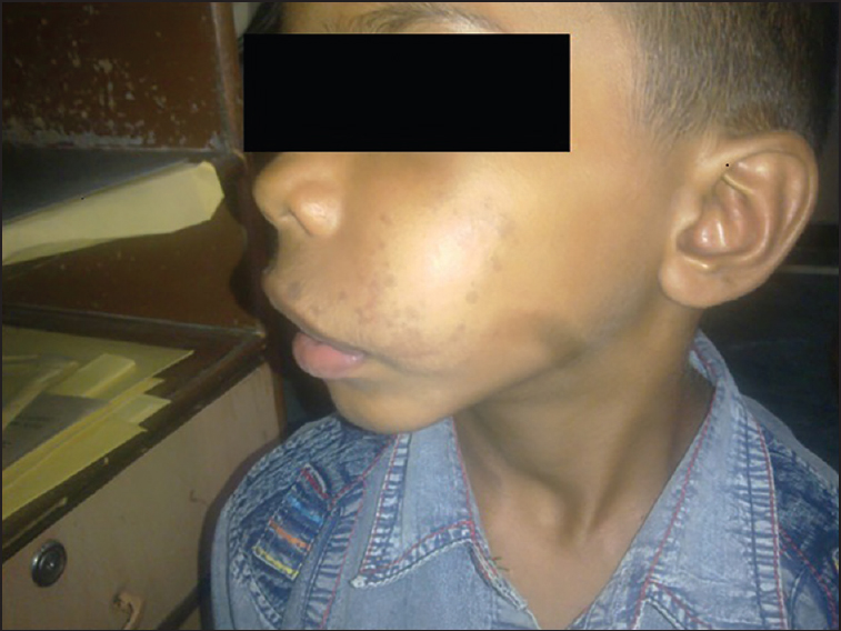
A 4-year-old boy presented with a diffuse swelling involving left cheek and left side of upper lip since birth causing disfigurement of his face. The diffuse lump started as a small red nodule at birth which gradually increased to the present size with the growth of the child. There was no history of any pain or bleeding from the lump. On examination, there was a diffuse, ill defined, soft, non-compressible swelling in the left cheek and left side of the upper lip [Figure 1]. Hyperpigmented spots were also noted on the skin of the left cheek. There was no bruit on auscultation.

- Large diffuse swelling seen on the left lower face involving left cheek and upper lip
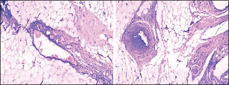
Doppler ultrasound was done which revealed a vascular lesion with doubtful communication with left facial vein. In view of the clinical presentation and inconclusive doppler report, a possibility of hemangioma followed by arteriovenous malformation was kept. Propanolol was started at the dose of 0.5 mg/kg and increased to 2 mg/kg but the swelling persisted. The child also received three cycles of intralesional bleomycin. However, there was no improvement in the size of the lump, despite treatment for 8 months. Finally, surgery by intraoral route was planned. The aim of the surgery was to explore the cheek and lip and attempt debulking. During surgery, it was found that there was dense fibrosis in the cheek with infiltration into underlying tissue and muscle resulting in difficult excision. The upper lip was also partially debulked and sent for histopathology. Microscopically, haphazard proliferation of capillary-sized vessels were seen mostly adjacent to vein wall along with abundant adipose tissue. Thick-walled larger blood vessels were also seen. At places these capillaries were arranged in a lobular pattern infiltrating into fat and adjoining muscle. Numerous nerve bundles, few of them showing myxoid change were also seen [Figure 2]. However, no arteries or arterioles were seen and vessel wall was negative for elastic tissue (Verhoffs stain). Based on the clinical details and histopathological findings, a final diagnosis of angiomatosis was given.

- Photomicrographs showing clusters of capillaries adjacent to vein wall infiltrating the adipose tissue with admixture of thick walled blood vessel, mature adipose tissue and nerve bundles. (Haematoxylin & Eosin stain, 400 x)
DISCUSSION
Vascular lesions especially hemangiomas are very common in childhood. The diagnosis and management of vascular lesions continue to pose a challenge for the histopathologists as well as the treating surgeon. Angiomatosis can be described as a diffuse form of hemangioma. However, it differs from hemangioma in its infiltrative nature and propensity for local recurrence. Hence the correct histopathological diagnosis is very important.
Rao and Weiss have suggested that the term ‘angiomatosis’ be used to connote a histologically benign vascular lesion that extensively involves a region of the body or several different tissue types in a contiguous fashion.[1] Angiomatosis is a histologically benign vascular lesion which involves a region of the body or several different tissue types in a contiguous fashion. Lesions reported radiologically as vascular malformation may in some instances correspond to what pathologists term angiomatosis.
Angiomatosis can be congenital or acquired. Acquired angiomatosis can be infectious due to HIV or Bartonellosis. Congenital form maybe sporadic or seen in association with certain syndromes such as Klippel Trenaunay syndrome, Sneddons syndrome or Gorham disease.[3] Kuffer et al. reported the first documented case of Klippel Trenaunay syndrome associated with visceral angiomatosis (gastrointestinal and genitourinary).[4] However, in our case no associated features of any syndrome were seen.
Angiomatosis usually presents in the first two decades of life mostly in childhood or adolescence with a slight predilection for females. Our patient was a 4-year-old male child. Howat et al.[5] reported 17 cases of angiomatosis presenting in children with recurrence seen in 10 patients and multiple recurrences in four patients.
This condition is usually seen in lower extremities. Very few cases are reported from head and neck region.[2] We present a rare case of angiomatosis in the face (cheek and lip). Angiomatosis has also been reported from other sites such as heart,[3] abdominal wall,[6] forearm,[7] retroperitoneum and genitalia.[4] Clinically, it mimics hemangioma or vascular malformation. In our case also the clinical diagnosis was hemangioma. The latter usually involutes by 2 year of age but vascular malformation grows persistently with age and does not disappear. As our patient did not respond to the bleomycin treatment for hemangioma, excisional biopsy and subsequent histopathological examination was planned.
Histopathologically, two types of patterns are seen in angiomatosis. The most common pattern consist of haphazard proliferation of varying sized vessels and clusters of capillary vessels adjacent to vein walls. In second pattern, a central large vessel is surrounded by clusters of capillary-sized vessels arranged in nodules. However, a distinctive feature common to both types is presence of large amount of adipose tissue. In the present case, predominantly the first pattern was seen. Large amount of mature fat frequently accompanying the vascular elements seen in angiomatosis suggest that this lesion may possibly be a generalised mesenchymal proliferation rather than an exclusively vascular lesion.[8]
Histologically, this lesion needs to be differentiated from angiolipoma because of the coexistence of vascular proliferation and abundant fat. However, in angiolipoma proliferating vessels are usually concentrated at the periphery of intratumoral lobules of adipocytes and the lesion is quite well circumscribed.[8] Other differential diagnosis includes angiomyolipoma, infiltrating lipoma, angiomyxolipoma and liposarcoma. Angiomyolipoma can be differentiated from angiomatosis as it contains smooth muscle in addition to blood vessels and fat while angiomyxolipoma is distinguished by the presence of myxoid change in adipose tissue and absence of haphazard blood vessel proliferation.[9] Val Bernal et al. reported a case of soft tissue angiomatosis closely mimicking liposarcoma due to the presence of large mass with prominent myxoid adipose tissue.[7] Features such as mitotic figures, cellular atypia and presence of typical lipoblasts help to differentiate liposarcoma from angiomatosis as they are not seen in latter. Large amount of mature fat frequently accompanying the vascular elements seen in angiomatosis suggest that this lesion may possibly be a generalised mesenchymal proliferation rather than an exclusively vascular lesion.[10]
Complete resection is preferred in local angiomatosis while radiotherapy or interferon α2a is the treatment of choice in extensive angiomatosis.[5] However, about 90% of patients demonstrate local recurrences on follow up.[1]
Through this article, we wish to highlight in a young patient presenting with diffuse persistent vascular swelling of the head and neck region, a possibility of angiomatosis, though rare, should be considered. The diagnosis is made on histopathology showing vascular elements admixed with abundant adipose tissue. The correct histopathological identification of this uncommon entity and its differentiation from the more innocous vascular lesion, i.e., hemangioma is important, considering its high recurrence rate.
Source of Support: Nil.
Conflict of Interest: None declared.
REFERENCES
- Angiomatosis of soft tissue. An analysis of the histological features and clinical outcome in 51 cases. Am J Surg Pathol. 1992;16:764-71.
- [Google Scholar]
- Primary left ventricular angiomatosis: First description of a rare vascular tumor in the left heart. Int Heart J. 2006;47:469-74.
- [Google Scholar]
- Klippel-trenaunay syndrome, visceral angiomatosis and thrombocytopenia. J Pediatr Surg. 1968;3:65-72.
- [Google Scholar]
- Angiomatosis: A vascular malformation of infancy and childhood. Report of 17 cases. Pathology. 1987;19:377-82.
- [Google Scholar]
- Angiomatosis of the abdominal wall: Imaging findings in three adults. Radiology. 1994;193:543-5.
- [Google Scholar]
- Soft- tissue angiomatosis in adulthood: A case in the forearm showing a prominent myxoid adipose tissue component mimicking liposarcoma. Pathol Int. 2005;55:155-9.
- [Google Scholar]
- Angiomatosis: A case with metaplastic ossification. Am J Dermatopathol. 2009;31:367-9.
- [Google Scholar]
- Lesions of the oral cavity. In: Gnepp DR, ed. Diagnostic Surgical Pathology of the Head and Neck (1st ed). Philadelphia: WB Saunders Company; 2001. p. :266.
- [Google Scholar]
- Vascular myxolipoma (“angiomyxolipoma”) of the spermatic cord. Am J Surg Pathol. 1996;20:1145-8.
- [Google Scholar]


