Translate this page into:
Nail Photography: Tips and Tricks
Address for correspondence: Dr. Feroze Kaliyadan, Department of Dermatology, King Faisal University, Al-Ahsa, Saudi Arabia. E-mail: ferozkal@hotmail.com
This is an open access article distributed under the terms of the Creative Commons Attribution-NonCommercial-ShareAlike 3.0 License, which allows others to remix, tweak, and build upon the work non-commercially, as long as the author is credited and the new creations are licensed under the identical terms.
This article was originally published by Medknow Publications & Media Pvt Ltd and was migrated to Scientific Scholar after the change of Publisher.
Abstract
Photographic documentation of the nails is important in the objective evaluation of response to treatment and in disseminating scientific information related to nail diseases. The key to a good image of the nail is proper framing and achieving a sharp focused image with good contrast with the background, at the same time avoiding strong reflections from the nail surface. While the general principles of clinical photography apply to nail imaging also, this article attempts to highlight some tips which can be specifically used to improve the quality of nail images.
Keywords
Clinical photography
imaging
nail
INTRODUCTION
Nail diseases are a common part of the dermatology spectrum. Photographic documentation of nail diseases is a useful part of objective evaluation of response to treatment. Good photographic documentation is also essential for proper dissemination of scientific knowledge in the form of presentations or publications.
GENERAL PRINCIPLES OF CLINICAL PHOTOGRAPHY
The basic principles of clinical photography remain the same for nail photography. Some of the important points to remember are as follows:
-
Taking patient consent
-
Proper framing – keep the area of interest centred and crop out blank space around the lesion as much as possible
-
Standardisation of images (especially for pre- and post-photographs) – use a tripod whenever possible, use uniform distances, angles and lighting for serial images
-
Take multiple images. Take images from different angles. You can always delete the poor images
-
Optimum lighting – This is probably the most crucial element of photography in general
-
Uniform, non-reflective background, preferably with a darker colour.[1]
SO WHAT IS DIFFERENT IN NAIL PHOTOGRAPHY?
The nail covers a relatively small area. In an ideal setup, a single-lens reflex camera with a dedicated macro lens would produce the best images. A ring light will be a useful additional tool as it would help to avoid strong reflections from the nail surface. The best way to capture the image would be to go back a bit and zoom out to capture the nails. This would help prevent the shape distortion which is associated with shooting from too close a distance. The same can be done even with a point-and-shoot camera with sufficient optical zoom (also make it a point to use the macro mode on the point and shoot).
Unless you are familiar with manual settings, it is always better to use automatic settings with the flash on. A strong reflection from the nail surface can be avoided by using a ring flash, reflectors or dulling the flash by covering the flash surface with a dedicated flash diffuser or tissue paper [Figures 1 and 2]. This is very important as the nail plate tends to produce strong reflections which may whiten out important morphological features.

- Effect of flash on nails (a) without flash diffusion (b) after flash correction
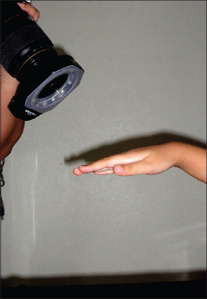
- Using dedicated ring flashes and macro lenses for nail photography
The background needs to be a uniform, non-reflective surface. A dark blue, black or green cloth is the best. However, to ensure good contrast between the nail and background, it is best to shoot the nails with a gap (about 2 m has been suggested) between the nails and the background, with the camera in the macro mode with flash on (’black background technique’).[2] We have found good results with a gap of even 5–10 cm between the nails and the background [Figures 3 and 4].
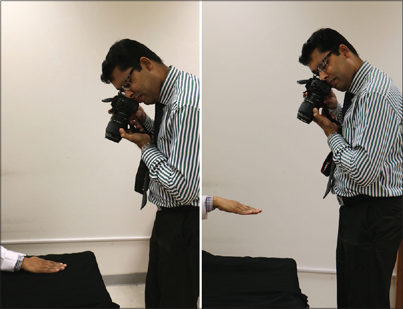
- Imaging of the nails with a dark non-reflective background with a gap between the nail and the background
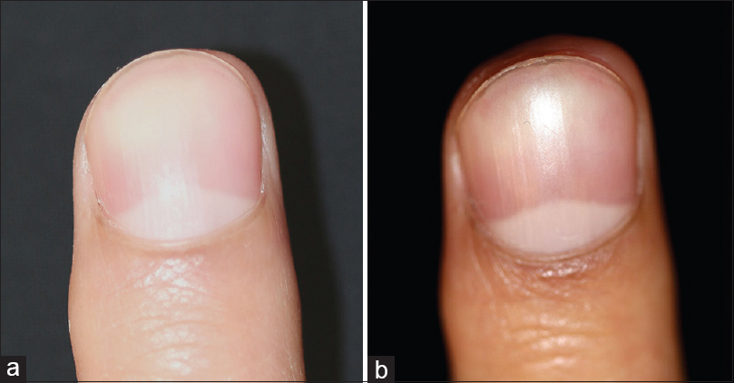
- Difference in the contrast illustrated between the images (a) nail placed directly on the background and (b) with a gap between the nail and the background
When the condition involves multiple nails, there are different techniques mentioned to include all nails in a single frame (or in minimum frames). Ashique and Kaliyadan described a technique, in which two sets of images are obtained; one including all fingernails except the thumb and a second image including only the thumb. The images are then combined into a collage [Figure 5]. This provides an image in which the nails are in the same plane, symmetrical and also looks aesthetically pleasing. Standardisation is also easier with this technique.[3] Gupta and Gupta suggested a modification of this method where the frame could be made less broad by placing one hand over the other [Figure 6].[4] Inamadar and Palit suggested a technique of imaging the nails in a position where the palmar aspects of both hands are kept side by side. Four fingers are flexed at proximal interphalangeal joints towards the palms. The thumbs are flexed across the palm and brought closer to the tips of other fingers in a way that the thumbnails face upwards [Figure 7]. The advantage of this technique is that all the nails can be included in a single frame. It is, however, difficult to standardise this technique and it may also pose difficulties in patients having joint problems, making flexion of the small joints difficult.[5]
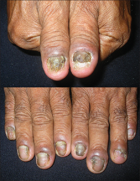
- Technique for placing all finger nails in a frame
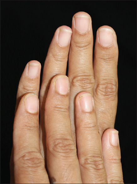
- Technique for placing all finger nails in a frame other than the thumb
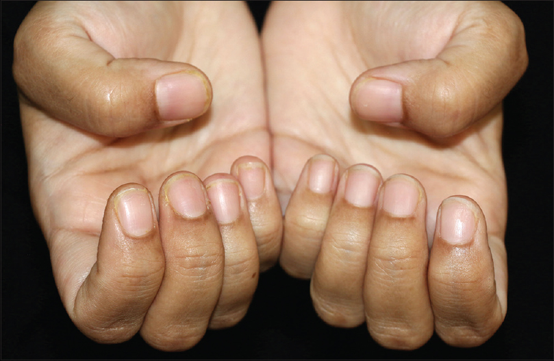
- Technique for placing all finger nails in a frame
MISCELLANEOUS
Onychoscopy (dermoscopy of the nails) is being increasingly used in enhancing the diagnosis of nail diseases. Both non-polarised, contact dermoscopy and polarised light dermoscopy can be used for the nails. Details regarding onychoscopy itself are beyond the scope of this article. However, we would like to highlight that documentation of dermoscopic images of the nails is sometimes difficult because of the curved convex surface of the nail. When using normal fluid contact dermoscopy, using a sufficiently thick amount of relatively firmer dermoscopic medium such as ultrasound gel is essential to fill the convex surface to get the best images with minimal peripheral blurring.[2]
Transillumination can sometimes be a useful tool in assessing the extent of subungual lesions such as wart. Since external light sources such as the camera flash will whiten out the transillumination, the image will have to be taken with the flash off [Figure 8]. Using a tripod would be essential to prevent camera shake as even a mild shake will blur details in the case.[6]
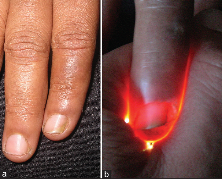
- (a) Normal lighting (b) Transillumination
PRACTICE POINTS
-
General principles of clinical photography apply to nail photography too
-
The ‘black-background’ or ‘dark-background’ technique, where the nails are photographed with a gap between it and a dark non-reflective background, gives the best results
-
The flash must be controlled to avoid excess reflections
-
Different standardised frames can be considered when all nails have to be placed in a single image
-
Consider using accessories such as macro lenses and ring flashes for the best results.
Financial support and sponsorship
Nil.
Conflicts of interest
There are no conflicts of interest.
REFERENCES
- Basic digital photography in dermatology. Indian J Dermatol Venereol Leprol. 2008;74:532-6.
- [Google Scholar]
- Baran R, de Berker DA, Holzberg M, Thomas L, eds. Imaging the nail unit. Baran and Dawber's Diseases of the Nails and Their Management (4th ed). Oxford: Wiley-Blackwell; 2012. p. :101-82.
- Clinical photography of nail diseases: A simple method to include all fingernails in a frame. J Am Acad Dermatol. 2015;72:e25.
- [Google Scholar]
- Transillumination: A simple tool to assess subungual extension in periungual warts. Indian Dermatol Online J. 2013;4:131-2.
- [Google Scholar]


