Translate this page into:
Successful Management of Dowling-Degos Disease with Combination of Q-switched Nd: YAG and Fractional Carbon Dioxide Laser
Address for correspondence: Dr. Swagata Arvind Tambe, Department of Dermatology, Topiwala National Medical College and BYL Nair Hospital, Mumbai, Maharashtra, India. E-mail: swagatatambe@gmail.com
This is an open access article distributed under the terms of the Creative Commons Attribution-NonCommercial-ShareAlike 3.0 License, which allows others to remix, tweak, and build upon the work non-commercially, as long as the author is credited and the new creations are licensed under the identical terms.
This article was originally published by Medknow Publications & Media Pvt Ltd and was migrated to Scientific Scholar after the change of Publisher.
Dear Editor,
Dowling-Degos disease (DDD) is a rare, inherited disorder characterised by numerous, asymptomatic, small, round-pigmented macules over axillae and groins, face, neck, arms and trunk, scattered comedo-like lesions (dark dot follicles) and pitted acneiform scars. Various treatment modalities have been tried without much benefit. We report a case of 19-year-old female with DDD successfully treated with a combination of Q-switched Nd: YAG and fractional carbon dioxide (CO2) laser.
A 19-year-old unmarried female presented with asymptomatic pigmented spots on the face, flexural areas such as axillae, groins, antecubital fossae, popliteal fossae and dorsum of hands since 8 years. The major complaint of the patient was cosmetic disfigurement produced by the pigmented spots and pits on the face. None of the family members were affected similarly.
Cutaneous examination revealed multiple hyperpigmented macules with comedo-like papules over face [Figure 1a and b], forehead [Figure 1c] sides of the neck, axillae, antecubital [Figure 1d] and popliteal fossae, groins [Figure 1e] and dorsum of the hands [Figure 1f]. Multiple pitted scars were present on the face, upper chest and back.

- (a and b) Multiple hyperpigmented macules with comedo-like papules and pitted scars seen over cheeks, temples of right and left side of the face, (c) multiple hyperpigmented macules and pitted scars on forehead, (d) multiple hyperpigmented macules in both antecubital fossae, (e) multiple hyperpigmented macules in the groin, (f) multiple hyperpigmented macules on dorsum of the hand
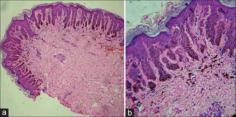
Skin biopsy from the hyperpigmented macules revealed hyperkeratosis, elongated and bifurcated rete ridges ('antler like' rete ridges) with increased melanin pigment at the lower part of rete [Figure 2a]. Keratotic plugging of the pilosebaceous orifice with melanin incontinence in the dermis [Figure 2b]. Based on clinical and histologic findings, diagnosis of DDD was made.

- (a) Skin biopsy from the hyperpigmented macules showing hyperkeratosis, elongated and bifurcated rete ridges (‘antler like’ rete ridges) with increased melanin pigment at the lower part of rete (H and E, ×40), (b) keratotic plugging of the pilosebaceous opening, elongated rete ridges with increased pigmentation of the rete ridges and melanin incontinence in the dermis (H and E, ×100)
The patient was advised laser therapy for her facial lesions and topical adapalene 0.1% for her body lesions. She was first treated with three sessions of Q-switched Nd: YAG laser every 3 weeks followed by two sessions of fractional CO2 laser each at an interval of 4 weeks. Depending on the lesions, for Q-switched Nd: YAG laser spot sizes used were ranging from 6 to 8 mm in diameter, with a frequency or repetition rate of 3–5 Hz and a pulse energy of 1000–1200 mJ (670–690 volts). Parameters used for fractional CO2 laser include spot diameter of 0.8–1.2 mm, pulse energy of 20–22 watts, pulse duration of 5–7 m, and spot density was 100–150 MTZ/cm2. Post-procedure sunscreen was advised, and topical adapalene was continued between the sessions.
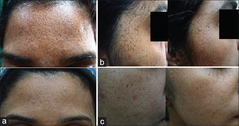
After 5 sessions of laser therapy, patient showed remarkable improvement in hyperpigmentation and scarring [Figure 3a–c]. The patient was followed up for 1 year with no recurrence of lesions.

- (a) Significant improvement in hyperpigmented macule, comedo-like papules and pitted scars on forehead, (b) significant improvement in hyperpigmented macules, comedo-like papules and pitted scars on the right temple and cheek, (c) significant improvement in hyperpigmented macules and pitted scars on the right cheek
DDD is a type of reticulate pigmentary disorder (OMIM Number: 179850). It is also known as reticulate pigmented anomaly of flexures and Dowling-Degos-Kitamura disease. The mode of inheritance is either sporadic or autosomal dominant. The proposed pathogenesis is a loss of function mutation on chromosome 12 (in the KRT5 gene encoding for keratin 5), leading to melanosome uptake deficiencies and structural defects in hair follicles and sebaceous glands.[1] The characteristic histologic feature is filiform elongation of rete ridges with antler-like configuration.[2] It usually appears after puberty with female preponderance. The sites commonly involved are axillae, groins, inframammary area, face, sides of the neck, popliteal and antecubital fossae, rarely genitals, vulva and back.[3] The major clinical manifestations are acquired hyperpigmentation affecting the flexures, pitted perioral acneiform scars and hyperkeratotic comedo-like lesions on the neck. Rare manifestations include dystrophic fingernails, multiple keratoacanthomas, pilonidal sinus, seborrheic keratosis and hidradenitis suppurativa.
Many different treatment options have been tried in recent years without convincing therapeutic benefits which includes depigmenting agents such as hydroquinone, as well as systemic and topical retinoids. Various lasers especially Erbium YAG and fractional Erbium YAG have been beneficial in treating DDD.[45]
Q-switched Nd: YAG laser 1064 nm has been successfully used in pigmentary disorders due to its longer wavelength, higher fluence and shorter pulse while it is less efficacious in scarring. Due to its selective photothermoloysis, there is no damage to the surrounding area. It is a painless procedure done in <20 min with mild erythema post-procedure which fades in an hour.
Fractional CO2 laser is effective in scarring due to its fractional photothermolysis and safe with continued improvement over time.
Combination of ablative technology with fractional thermolysis is an effective option for pigmentation and scarring with minimal side effects. This combination was successfully used in acne scarring and exogenous ochronosis.
Considering the limited treatment options, our case suggests that combination of Q-switched Nd: YAG and fractional (CO2) lasers might be a successful strategy in the management of DDD.
Financial support and sponsorship
Nil.
Conflicts of interest
There are no conflicts of interest.
REFERENCES
- Loss-of-function mutations in the keratin 5 gene lead to Dowling-Degos disease. Am J Hum Genet. 2006;78:510-9.
- [Google Scholar]
- Dowling-Degos disease (reticulate pigmented anomaly of the flexures): A clinical and histopathologic study of 6 cases. J Am Acad Dermatol. 1999;40:462-7.
- [Google Scholar]
- Dowling-Degos disease involving the vulva and back: Case report and review of the literature. Dermatol Online J. 2011;17:1.
- [Google Scholar]
- Successful treatment of Dowling-Degos disease with Er: YAG laser. Dermatol Surg. 2002;28:748-50.
- [Google Scholar]
- Treatment of Dowling-Degos disease with fractional Er: YAG laser. J Cosmet Laser Ther. 2013;15:336-9.
- [Google Scholar]


