Translate this page into:
Calculating Area of Graft Required for Vitiliginous Areas During Split-thickness Skin Grafting: A Simple, Accurate, and Cost-effective Technique
Address for correspondence: Dr. Tasleem Arif, New Colony Soura, near water supply control Room, Srinagar, Kashmir 190011, India. E-mail: dr_tasleem_arif@yahoo.com
This is an open access article distributed under the terms of the Creative Commons Attribution-NonCommercial-ShareAlike 3.0 License, which allows others to remix, tweak, and build upon the work non-commercially, as long as the author is credited and the new creations are licensed under the identical terms.
This article was originally published by Medknow Publications & Media Pvt Ltd and was migrated to Scientific Scholar after the change of Publisher.
Sir,
Vitiligo is an acquired disorder of pigmentation affecting approximately 1% of the world’s population and is characterized by depigmented macules with a significant impact on the quality of life.[1] Various surgical procedures have been used for resistant, stable vitiligo not responding to medical or light therapy. Split-thickness skin grafting involves the transplantation of a sheet of epidermis with variable amount of dermis to the dermabraded vitiliginous area. The method to calculate an absolute area of graft required for repigmenting the vitiliginous area has not been standardized so far as there is no universally designed technique for calculating the required area of graft for vitiligo surgery. In this article, the authors describe a technique that is accurate, simple, and cost-effective to measure the area of vitiliginous lesion/s, which will guide the dermatosurgeon for harvesting the required amount of graft ensuring no wastage of graft or avoiding the risk of inadequate harvest of graft.
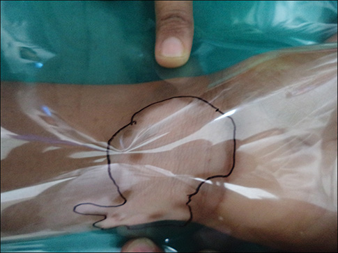
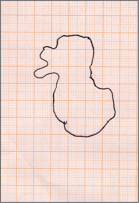
Technique: The technique is very simple and relies on simple mathematics. The surgeon needs a graph paper (or an ECG paper), a transparent plastic film (plastic transparent sheets used for overhead projector), and a marking pen. Put the transparent film over vitiligo lesion/s. With the help of a sketch pen, mark the boundaries of lesion/s over the film [Figure 1]. Put this marked film over a graph paper [Figure 2] and secure with staples so that the marked sheet does not move here and there to affect readings of calculated area.

- A vitiligo lesion is covered by a transparent plastic sheet and marked over with a sketch pen. The borders of the lesion on the skin can also be marked before putting the film over it in case borders are not well defined

- Scanned picture with marked transparent film kept over the graph paper
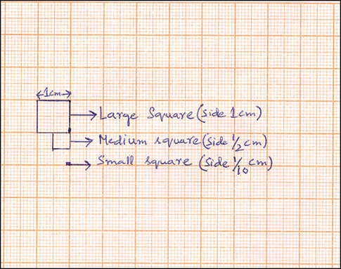
Calibration of graph paper: A graph paper has two types of squares. The sides of the large square measure 1 cm [Figure 3]. Each large square is composed of 100 small squares so that the side of each small square is 1 mm (1/10 cm). Thus, the area of one large square is (1 cm × 1 cm = 1 cm2) 1 cm2 and the area of one small square is 1 mm2 or 1/100 cm2 (1 mm × 1 mm = 1 mm2 or 1/10 cm × 1/10 cm = 1/100 cm2).

- Calibrations shown on the graph paper. A large square has side 1 cm. Each large square is composed of 100 small squares so that the side of each small square is 1 mm (1/10 cm). One medium square corresponds to 25 small squares or ½ cm side
Calculation of area for grafting: Under the marked area, count the number of complete large squares. At some places, the marked area involves half, less than half, or more than half of large squares. For them, count number of small squares. The total area in cm2 required for grafting is given by the following formula:
Area required for grafting (in cm2)= No. of complete big squares + 1/100 X No. of small squares
For example, a vitiligo lesion kept for grafting, on measuring, consisted of complete 7 big squares and 170 small squares. The area of graft required is as follows:
Area(in cm2)=7+1/100×170=7+1.7=8.7 cm2
Thus, depending on the number of lesions to be treated surgically, individual lesional scores can be combined to give the total area of graft required for the surgery.
How to count small squares at a faster rate?—Modification of the technique: Many surgeons may find it a bit laborsome to count small squares. To hasten the calculation, some modifications are made. By looking precisely at a graph paper, a large square is found to be divided into four medium squares [Figure 3] by two relatively bold lines, one midway vertical and another horizontal such that one medium square corresponds to 25 small squares or ¼ cm2 area. Thus, formula is modified as follows:
Area (in cm2) = No of complete big squares + 1/4 × No. of complete medium squares + 1/100 × No. of small squares.
For example, a vitiligo lesion on measuring was found to be having 8 complete big squares, 4 medium squares, and 35 small squares. Area of skin graft required is as follows:
Area (in cm2)= 8+1/4×4+1/100X35=8+1+0.35 =9.35 cm2
Advantages of the technique:
-
This technique is simple.
-
It is very accurate.
-
It is cost-effective as it does not involve any instrument.
-
All the requirements are readily available
-
It ensures an absolute measurement of graft area so that no excessive graft is harvested, thereby minimizing wastage.
-
It can also be used for monitoring vitiligo during treatment to assess pre- and posttreatment area.
-
It can be used for charting as well as scoring of other diseases such as melasma, leprosy, plaque psoriasis, scleroderma, etc.
At some centers, researchers follow a similar method to calculate the area of ultrathin skin grafting for leukoderma. A transparent film is put over the marked vitiligo areas and the markings of the lesion/s are transferred to the film. This film is then xeroxed using a copier machine and the outlined areas on the paper are cut out and then weighed on an electronic balance. The total area is then calculated depending on the weight of cut paper.[2] However, the present technique does not require a copier or a weighing balance. Also, the present technique gives area in absolute number, i.e., in cm2. Thus, authors conclude that the present technique can help surgeons to calculate the graft area required for split-thickness grafting for vitiligo. This can also help researchers to calculate disease areas used for scoring of various diseases.
REFERENCES
- Ultrathin split-thickness skin grafting followed by narrowband UVB therapy for stable vitiligo: An effective and cosmetically satisfying treatment option. Indian J Dermatol Venereol Leprol. 2012;78:159-64.
- [Google Scholar]
- Treatment of leukoderma by transplantation of ultra-thin epidermal sheets. In: Gupta S, Olsson MJ, Kanwar AJ, Ortonne JP, eds. Surgical management of vitiligo (1st ed). Massachusetts: Blackwell Publishing; 2007. p. :115-22.
- [Google Scholar]


