Translate this page into:
Radiofrequency-Assisted Body Piercing
Address for correspondence: Ravi Kumar Chittoria, Professor, Department of Plastic Surgery, Jawaharlal Institute of Postgraduate Medical Education and Research (JIPMER) Pondicherry, India. E-mail: drchittoria@yahoo.com
This is an open access article distributed under the terms of the Creative Commons Attribution-NonCommercial-ShareAlike 3.0 License, which allows others to remix, tweak, and build upon the work non-commercially, as long as the author is credited and the new creations are licensed under the identical terms.
This article was originally published by Medknow Publications & Media Pvt Ltd and was migrated to Scientific Scholar after the change of Publisher.
Abstract
The art of body piercing is ancient; however, nowadays it has evolved into a fashion statement. In the Indian subcontinent, ear and nose piercing hold religious and cultural significance in addition to being done for aesthetic reasons. Body piercing is routinely performed by railroading technique or by piercing guns; many modifications of the technique have emerged. Irrespective of the technique used, the main complications associated are intraoperative bleeding and postoperative infection. To overcome these problems, we describe a novel and simple technique of ear and nose piercing using the radio frequency cautery.
Keywords
Body piercing
radiofrequency
bloodless piercing
INTRODUCTION
Ear piercing in the Indian subcontinent is not just done as an aesthetic procedure but performed routinely for religious and cultural purposes. It is probably due to this reason that the technique of ear piercing is constantly evolving with many innovations coming up over the years. Traditional methods involved passing a heated gold or silver wire through the ear lobule to create a suitable sized tract, which was immediately replaced with a gold ornament. This technique was superseded by the use of ear piercing guns that ensured a faster and less painful procedure.[1] The most common technique performed by medical practitioners involves railroading the earring onto a suitable sized needle and withdrawing it into the track created by the needle. Irrespective of the technique used, intra-operative bleeding and post-operative perichondritis often complicate this simple procedure. To overcome these problems, we describe a novel technique of ear piercing using a radiofrequency cautery.
CASE REPORT
A 30-year-old female patient with no keloidal tendencies presented to the plastic surgery OPD for ear and nose piercing. The patient already had an ear piercing on the lobule and wanted two additional piercings made above the existing one. The site for piercing was marked and sterile preparation of the ears and nose was done. A topical anesthetic agent containing a mixture of lignocaine and prilocaine (Eutectic mixture of local anesthesia) was applied under occlusion on the skin 2 hours prior to surgery. After ensuring adequate anesthesia a monopolar radiofrequency probe with a sterile pointed tip needle electrode was used to pierce the ear at the desired point [Figure 1]. The radiofrequency unit (Surgitron® FFPF EMC) was set on a cutting and coagulation (rectified) mode with a power dial of 2 [Figure 2]. A 18G needle was threaded over the radiofrequency electrode and brought out anteriorly from behind the ear [Figure 3]. The earring was secured over the tip of the 18G needle and pulled into the track created while withdrawing the needle [Figure 4].
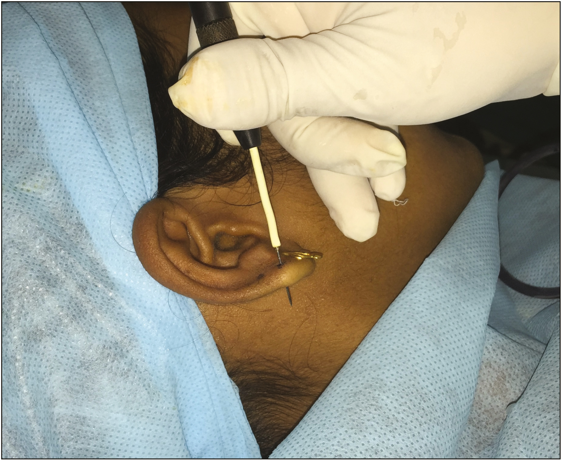
- Radiofrequency electrode used for piercing
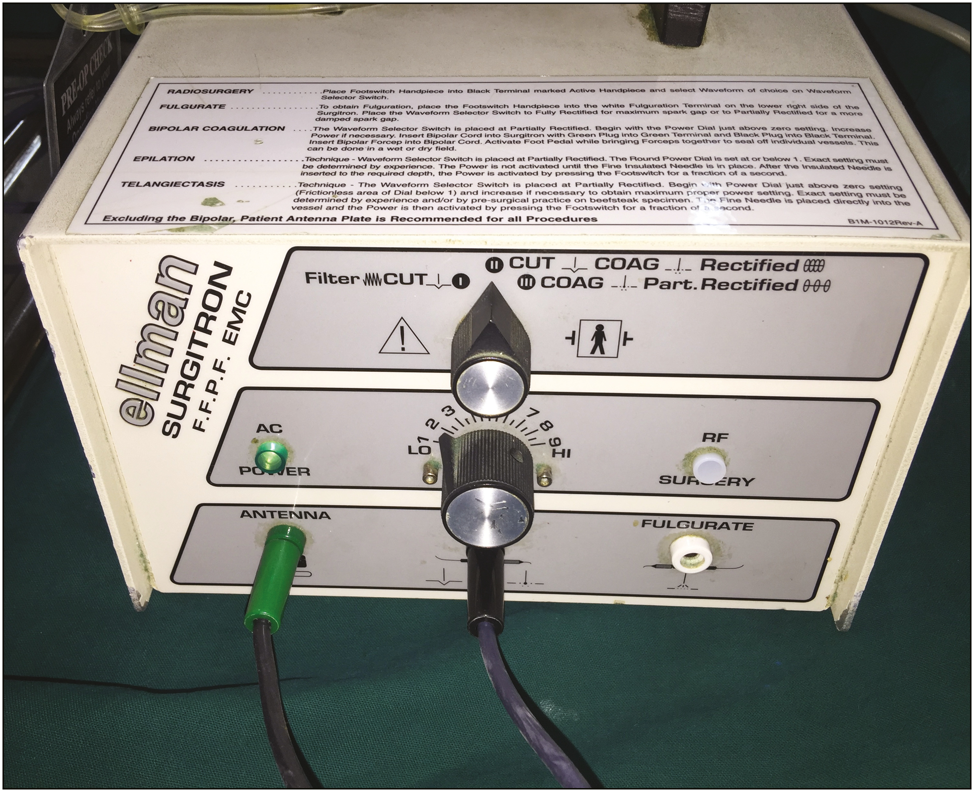
- Radiofrequency unit
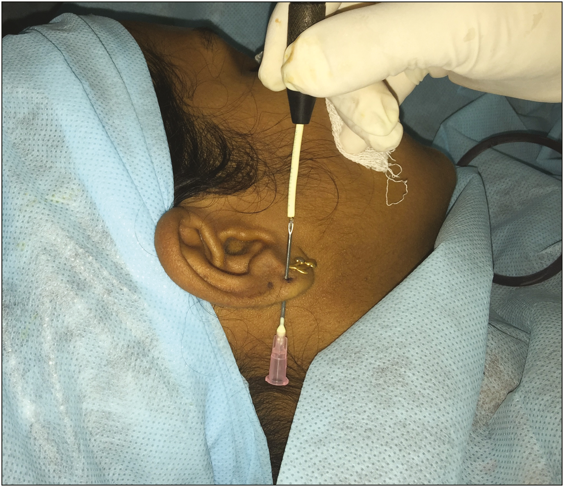
- 18G needle threaded over the radiofrequency electrode
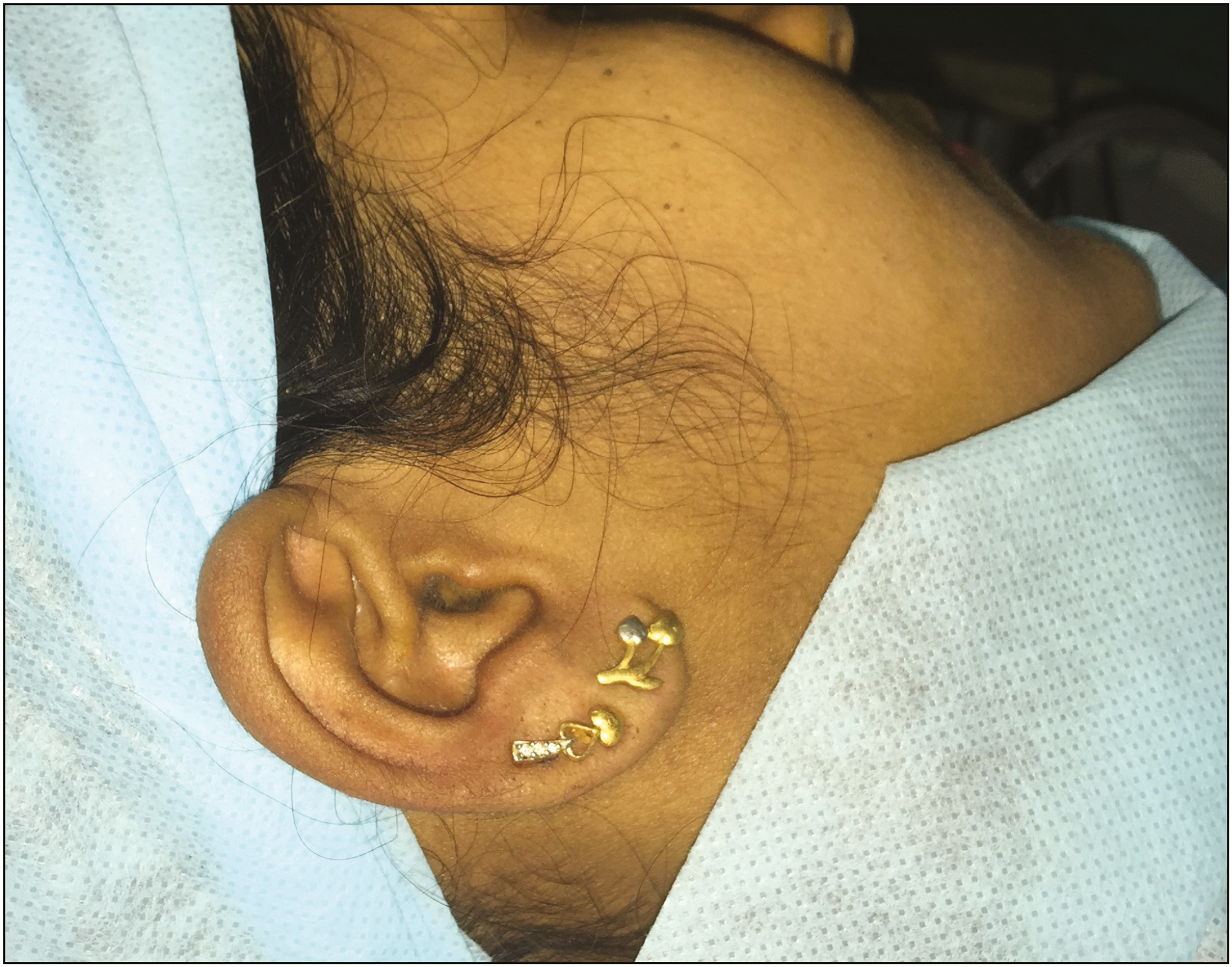
- Earrings in place
The second piercing was done similarly at a higher point, followed by piercing of the other ear and the nose [Figures 5 and 6]. Intraoperatively, the procedure was painless for the patient and there was only minimal bleeding. The surrounding skin sustained no damage. Local application of mupirocin ointment was done. No other oral antibiotics were administered. The patient was followed up for a week and no postoperative infection was observed.
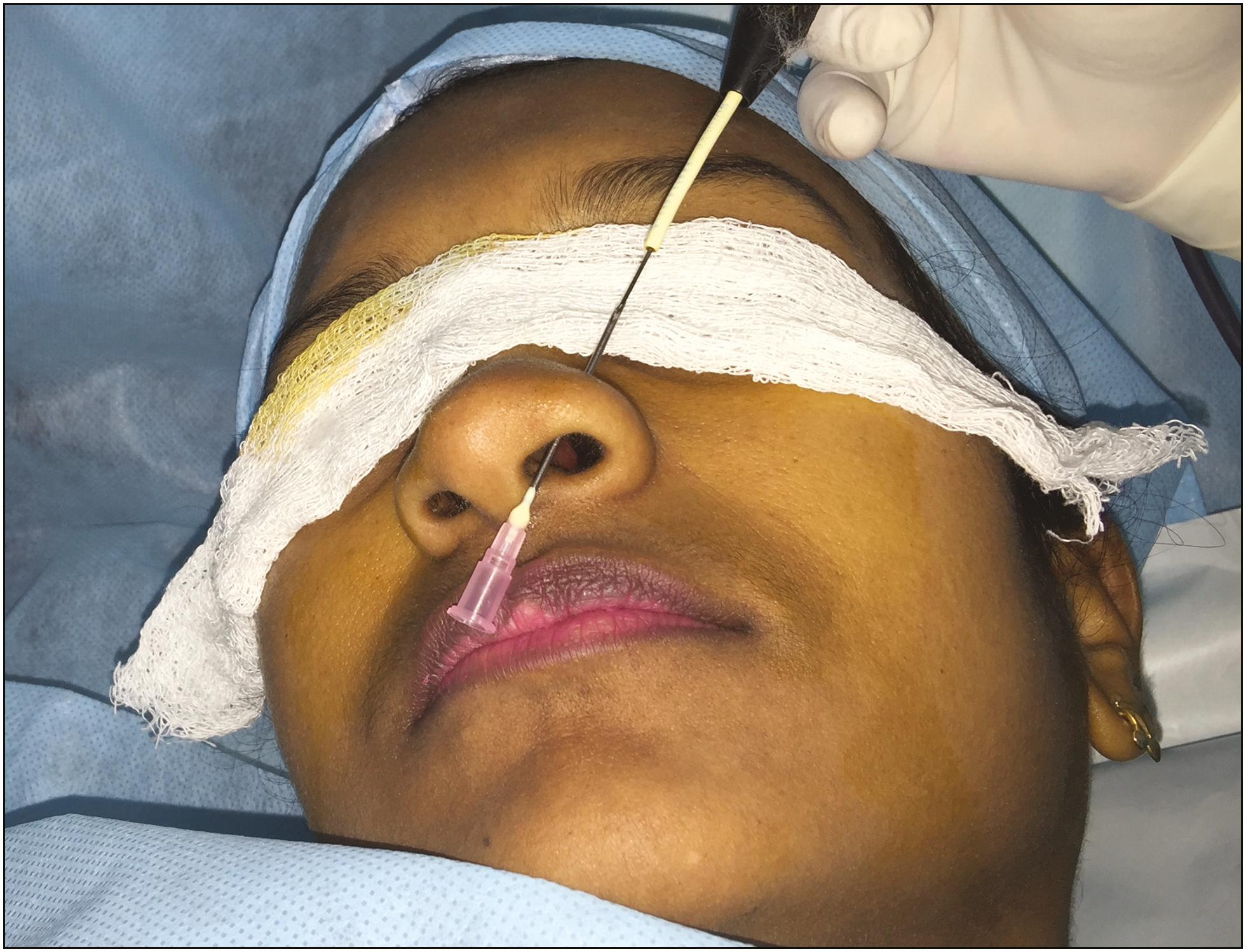
- Piercing done on the nose
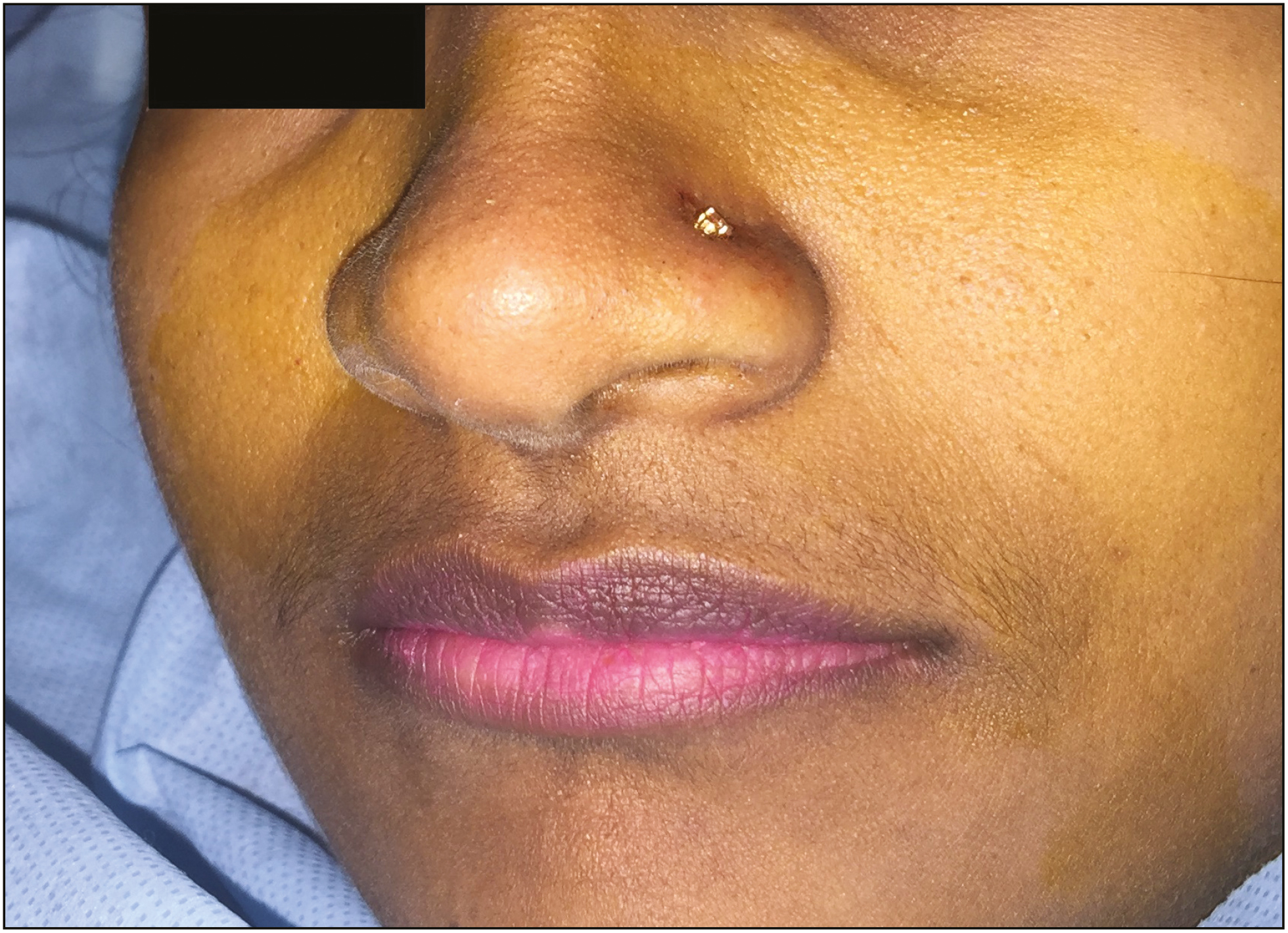
- Nose pin in situ
DISCUSSION
Radiofrequency has found widespread use in cosmetic surgery. It is frequently used in the ablation of nevi, vascular lesions, infective lesions such as warts, and also for facial rejuvenation and body contouring.[2] The radiofrequency electrode remains cold during the entire procedure and only the tip of the electrode that comes in contact with the tissue conducts the radiofrequency waves. As the diameter of the tip of the electrode is small, the electrode-tissue interface is small. This causes minimal collateral damage to the tissues (up to 75 mm).[3] In addition, the radiofrequency electrode can cut and coagulate at the same time ensuring good hemostasis during the surgery. This enables surgery to be performed without applying clamps on the earlobe or without injecting lignocaine adrenaline mixture into the operative site. The radiofrequency unit that was used in the above case was the Surgitron® FFPC EMC which has a 90 Watt Radiofrequency generator with four distinct waveforms; fully filtered, fully rectified, partially rectified, and fulguration.
Van Wijk et al.[4] in their study on human cadaver ears, have shown that despite the popular belief, both techniques, i.e. needle piercing and piercing guns result in almost equal damage to the cartilage. They also concluded that postoperative care and hygiene should be the focus to prevent perichondritis. In vitro studies done on cartilage have shown that after the use of radiofrequency, no alteration in the nuclear cytoplasm or lacunae were noted compared to the untreated areas.[5] Thus, radiofrequency probe seems to be safer to use on cartilage bearing areas of the ear and on the nose. Although the radiofrequency cautery is safe, rare complications such as pain, tissue edema, post inflammatory pigmentation changes, scarring and keloid formation can occur.[6]
The major shortcoming of our study is the lack of histological evidence to prove the lack of damage of radiofrequency on cartilage. As our observation is based on a single case report, further studies with histopathological correlation would be necessary to validate the same.
CONCLUSION
Radiofrequency technology is a safe, efficacious method for body piercing. After topical anesthesia, it provides painless and almost bloodless piercing. As hemostasis is maintained throughout the procedure, the piercing can be done quickly, preventing damage to the underlying cartilage or the surrounding skin.
Declaration of patient consent
The authors certify that they have obtained all appropriate patient consent forms. In the form the patient(s) has/have given his/her/their consent for his/her/their images and other clinical information to be reported in the journal. The patients understand that their names and initials will not be published and due efforts will be made to conceal their identity, but anonymity cannot be guaranteed.
REFERENCES
- A novel technique for piercing of ear lobule suited to Indian subcontinent. Indian J Plast Surg. 2013;46:160-1.
- [Google Scholar]
- Electrosurgery principles: Cutting current and cutaneous surgery–part I. J Dermatol Surg Oncol. 1988;14:29-31.
- [Google Scholar]
- Ear piercing techniques and their effect on cartilage, a histologic study. J Plast Reconstr Aesthet Surg. 2008;61(Suppl 1):S104-9.
- [Google Scholar]
- The acute effects of radiofrequency energy in articular cartilage: An in vitro study. Arthroscopy. 2000;16:2-5.
- [Google Scholar]


