Translate this page into:
Surgical Management of Onychoheterotopia
Address for correspondence: Dr. Chander Grover, Department of Dermatology and STD, Dilshad Garden, Delhi-110095, India. E-mail: chandergroverkubba@rediffmail.com
This is an open access journal, and articles are distributed under the terms of the Creative Commons Attribution-NonCommercial-ShareAlike 4.0 License, which allows others to remix, tweak, and build upon the work non-commercially, as long as appropriate credit is given and the new creations are licensed under the identical terms.
This article was originally published by Medknow Publications & Media Pvt Ltd and was migrated to Scientific Scholar after the change of Publisher.
Abstract
Onychoheterotopia (ectopic nail) is a rare condition characterized by the development of nail tissue, distinct from the normal nail unit. It is usually acquired following traumatic inoculation of nail matrix; the congenital variety being less common. The exact pathogenesis of the disease is not clear. It affects the dorsal aspect of fingers and toes mostly. Herein, we report a case of a 35-year-old man with post-traumatic onychoheterotopia of left middle finger, who was treated with surgical avulsion of the ectopic nail along with chemical matricectomy of the well-formed ectopic matrix. The patient had a satisfactory cosmetic outcome with normal growth of the nail unit and no recurrence. The report serves to highlight clinical presentation of acquired onychoheterotopia along with its surgical management.
Keywords
Chemical matricectomy
ectopic nail
phenol
INTRODUCTION
Onychoheterotopia (ectopic nail) is defined as continuous growth of nail tissue at any site other than the nail unit.[1] It is a rare entity that is usually acquired; the congenital variety being less common.[2] The exact pathogenesis is unknown, although various hypotheses have been proposed regarding its origin. Histopathologically, onychoheterotopia comprises a mature nail unit, including a cornified nail plate and nail matrix.[2] Partial excision, leaving behind the ectopic nail matrix, is frequently associated with recurrences.
CASE REPORT
A 35-year-old man, who was a farmer, presented with an extra nail plate over the left middle finger following a sharp injury at the same site 6 years back. Three months following the injury, he had noticed a nail-like outgrowth projecting from the proximal nail fold (PNF), which slowly grew just like the underlying nail plate, but was much thicker and deformed. This ectopic nail produced discomfort while working, although there was no pain or swelling.
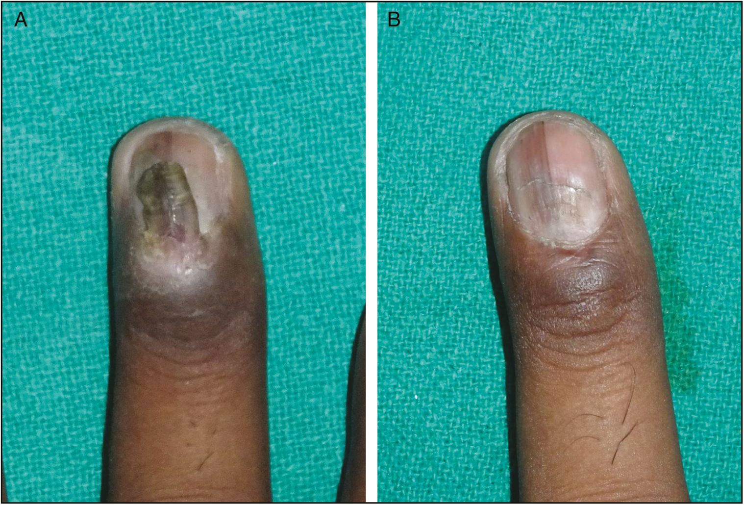
On physical examination, a well-defined nail plate distinct from the classical nail plate was found to arise from the PNF. It was rough and pigmented. The underlying normal nail plate showed a linear depression and an altered curvature, corresponding to the overlying ectopic nail plate [Figure 1A]. No bony deformity was apparent.

- (A) A deformed ectopic nail arising over a normal nail plate. It was arising from a well-formed pocket within the PNF. The ectopic nail shows discoloration and the underlying plate shows a slight depression. (B) Postoperative outcome at the end of 8 weeks, with a normal but slightly retracted PNF. Note the slow normalization of the nail plate curvature
Dermatoscopic examination of the nail (onychoscopy) showed an ectopic nail structure, distinct from the normal underlying nail plate, with an evidence of hematoma within its structure. It was arising from its very own, well-formed PNF with normal nail fold capillaries; however, the cuticle was absent. This indicated that the ectopic nail matrix was probably situated in an invaginated pocket under the dorsal layer of the PNF.
A clinical diagnosis of acquired onychoheterotopia was made. As the ectopic matrix was seemingly placed underneath the dorsal layer of the PNF, we planned to avulse the ectopic nail plate with total matricectomy of the ectopic matrix. The procedure was explained to the patient and a written informed consent was obtained.
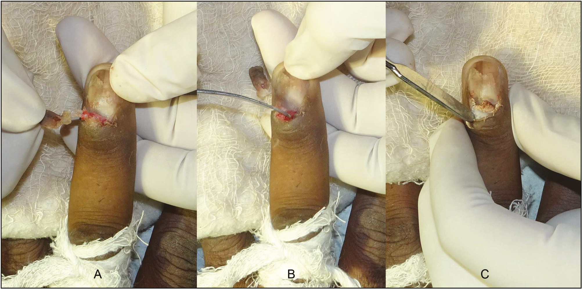
Under aseptic conditions, a proximal digital block was administered using 2% lignocaine without adrenaline. The digit was then exsanguinated and tourniquet was tied at the base of the digit to achieve an avascular operating field. The ectopic nail was avulsed [Figure 2A], exposing the ectopic matrix [Figure 2B]. This was followed by total matricectomy of this ectopic germinal matrix with 88% phenol using cotton-tipped applicators (1min of application time) to ensure complete destruction of the displaced nail matrix [Figure 2C]. The wound was then covered with nonadherent dressing. Postoperatively, oral antibiotics and analgesics were prescribed. The wound healed with a cosmetically acceptable outcome [Figure 1B]. Histopathological examination revealed a well-formed nail plate with germinal matrix without a granular layer. No recurrence or spicule formation was reported over a further 6-month follow-up period.

- Steps of surgery. After proximal digital block, exsanguination, and tourniquet, the ectopic nail was completely avulsed (A). This exposed the matrix from which the nail plate was arising (B). Redundant tissue was surgically removed and matricectomy was done with 88% phenol applied for 1min within the PNF pocket (C)
DISCUSSION
Onychoheterotopia is usually an acquired condition, though congenital cases can also be seen. It commonly follows a traumatic inoculation of nail matrix. The exact pathogenesis is unknown. Various hypotheses that have been proposed include ectopic existence of germ cells, nail of a rudimentary polydactyly, or traumatic splitting and implantation of germinal matrix at an ectopic site following trauma.[13] Various reports have shown association with palmar nail syndrome, Pierre Robin syndrome, and aberration of long arm of chromosome 6. This indicates a possible genetic predisposition.[4] Such cases are usually associated with an underlying bony anomaly. Congenital onychoheterotopia is most often seen on the palmar aspect of fifth digit[1]; whereas post-traumatic form is mostly seen on the dorsal aspect of the affected digit.[1] Presence at an extra-digital site is rarer but reported.
Onychoheterotopia may present as a small outgrowth of a deviant nail or a complete double finger nail malformation. It is usually asymptomatic but can be associated with pain, pruritus, infection, or impingement onto the matrix interfering with normal nail growth.[1]
Various patterns of growth have been described for ectopic nail. These include a horizontal nail (simulating a normal nail), vertical nail, or a circumferential nail.[1] Our case had a well-formed but dystrophic nail plate, placed over an otherwise normal nail plate. An ectopic nail is mostly deformed, probably, because of the absence of normal nail folds and nail bed, both of which have a favorable modulating influence on nail plate formation.[5] At times, if the ectopic nail matrix comes in contact with the periosteum, it may impede intramembranous ossification, producing hypoplasia and thinning of the phalanx.[6] Our patient had a normal underlying bone, presumably, because the ectopic matrix was placed distant from the phalanx.
Differential diagnoses to be considered for an ectopic nail include rudimentary polydactyly, cutaneous horn, foreign body, hamartoma, and split-nail deformity.[1] Histopathology offers confirmatory evidence if there is the presence of a fully developed nail unit, including nail plate and nail matrix. The presence of a nail bed is not necessary.[7] Both of these were confirmed in our patient.
Regardless of the type of onychoheterotopia, the standard treatment is surgical excision of the ectopic nail including the ectopic matrix, followed by primary closure of the defect. We resorted to the same in our patient who was much distressed as this ectopic appendage interfered with his handling of tools. A large defect may require flap reconstruction.[4] Fortunately in our case, ectopic matrix was superficially placed as well as adequately exposed after nail avulsion, enabling chemical matricectomy and simultaneous reconstruction of the PNF cover over the normal nail plate. The PNF has an important role in ensuring a shiny surface of the nail plate as well as the formation of the cuticle; thus, it was important to preserve it completely. At times, a partial matricectomy may result in a recurrence of ectopic nail, even if in the form of spicules. Our case had no such recurrence over the ensuing 6-month period.
CONCLUSION
Complete avulsion of the ectopic nail along with total ectopic matricectomy, while preserving the normal nail matrix and PNF, is important for the prevention of recurrence and maintaining cosmesis in cases with ectopic nail. Our report serves to highlight the usefulness of this approach.
Declaration of patient consent
The authors certify that they have obtained all appropriate patient consent forms. In the form the patient(s) has/have given his/her/their consent for his/her/their images and other clinical information to be reported in the journal. The patients understand that their names and initials will not be published and due efforts will be made to conceal their identity, but anonymity cannot be guaranteed.
Financial support and sponsorship
Nil.
Conflicts of interest
There are no conflicts of interest.
REFERENCES
- Onychoheterotopia: pathogenesis, presentation, and management of ectopic nail. J Am Acad Dermatol. 2011;64:161-6.
- [Google Scholar]


