Translate this page into:
Impacted Foreign Bodies in the Maxillofacial Region–A Series of Three Cases
Address for correspondence: Dr. Pulkit Khandelwal, Department of Oral & Maxillofacial Surgery, Pacific Dental College and Hospital, Udaipur, Rajasthan, India. E-mail: khandelwal.pulkit22@gmail.com
This is an open access journal, and articles are distributed under the terms of the Creative Commons Attribution-NonCommercial-ShareAlike 4.0 License, which allows others to remix, tweak, and build upon the work non-commercially, as long as appropriate credit is given and the new creations are licensed under the identical terms.
This article was originally published by Medknow Publications & Media Pvt Ltd and was migrated to Scientific Scholar after the change of Publisher.
Abstract
Penetrating injuries to the maxillofacial region are very common. Foreign bodies embedded deep in the maxillofacial region due to these injuries pose a challenge to an oral and maxillofacial surgeon. These objects may become a potent source of pain and infection. Early diagnosis of these foreign bodies can be achieved by the use of plain radiographs, ultrasonography, computed tomographic scans, and magnetic resonance imaging. Once diagnosed and located, these foreign bodies should be removed. Here, we report three such cases where early diagnosis of these foreign bodies embedded in the maxillofacial region lead to their early and successful removal without complications.
Keywords
Foreign body
infection
maxillofacial
trauma
INTRODUCTION
In any penetrating injury in the head and neck region, the presence of foreign body should be considered. Type of object, size of object, anatomical proximity of the foreign body to vital structures, and difficulty in retrieving may present challenges to the surgeon. Pieces of metallic objects, broken wood, twigs, bamboo splinter, glass particles, tooth brush, fish hook, pen cap with spring, and fragments of smoking pipe are some foreign bodies that get impacted in the maxillofacial region.[1]
Among all cases of foreign body contamination or impaction, about one-third are missed during initial examination. Sometimes, these foreign bodies may remain dormant in the soft tissue for years without causing any significant damage to the adjacent structures. However, in most of the cases, the presence of foreign substance can result in acute or chronic inflammation causing persistent and distressing symptoms. Diagnosis of these cases is often due to the presence of associated pain and swelling. Sometimes, it is also accidental on radiographic examination.[23]
CASE 1
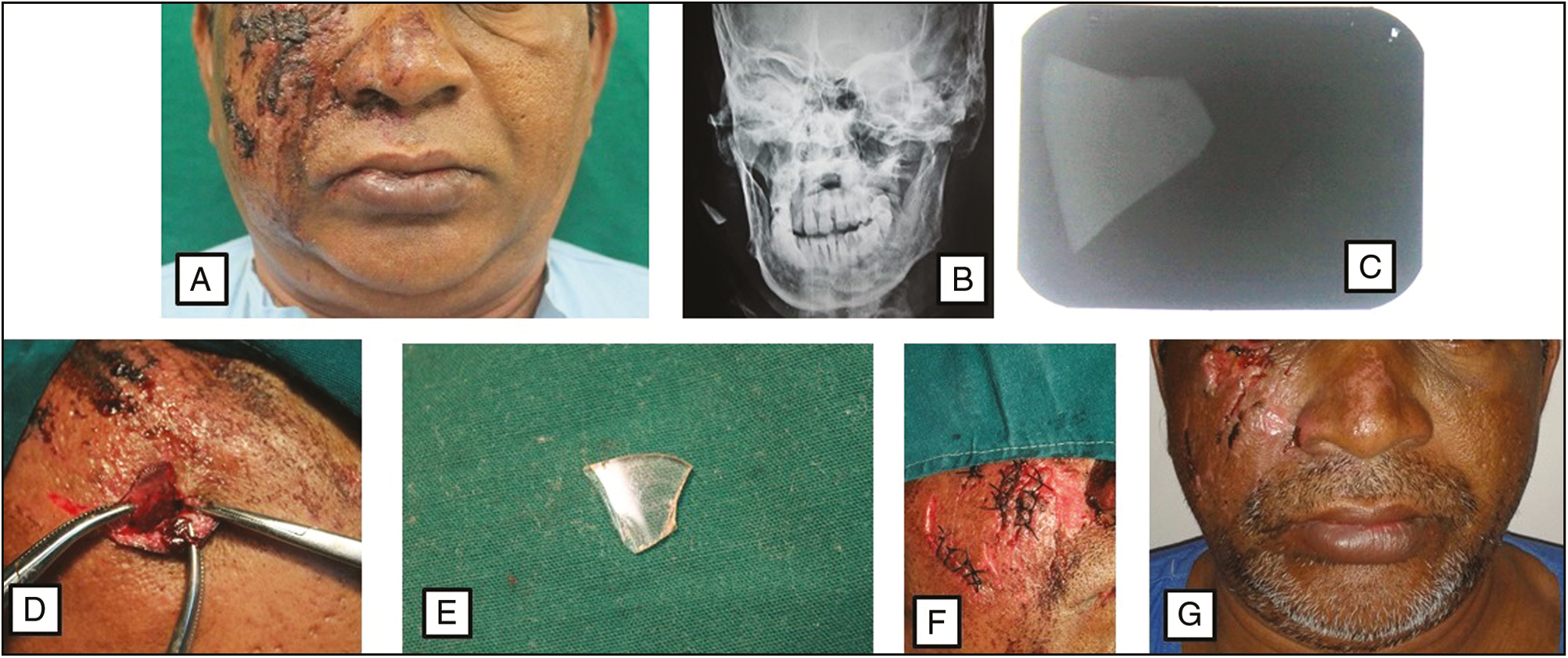
A 50-year-old male patient had a chief complaint of pus discharge from the cheek on the right side since 3 days. He had sustained injury over the right infraorbital and malar region in a road traffic accident a week earlier and treatment (suturing) was done at another hospital. On local examination, sutured lacerations and scabs were present over the right infraorbital and malar region [Figure 1A]. Pus discharge was present. Posteroanterior (PA) view was done which revealed a radio-opaque object at the level of mandibular ramus at occlusal level [Figure 1B]. The malar region was again palpated bimanually and it raised suspicion of some foreign body below the malar region just ahead of the ramus region. Intraoral periapical radiograph (IOPA) of the soft tissue cheek revealed a radio-opaque trapezoidal object [Figure 1C].

- (A) Preoperative view. (B) PA mandible confirming presence of a radio-opaque object in the right ramus area of the mandible. (C) IOPA confirming presence of a radio-opaque object in the right ramus area of the mandible. (D) Retrieval of the embedded foreign body through the existing wound. (E) Foreign body which turned out to be broken glass piece. (F) Closure. (G) One week follow up
CASE 2
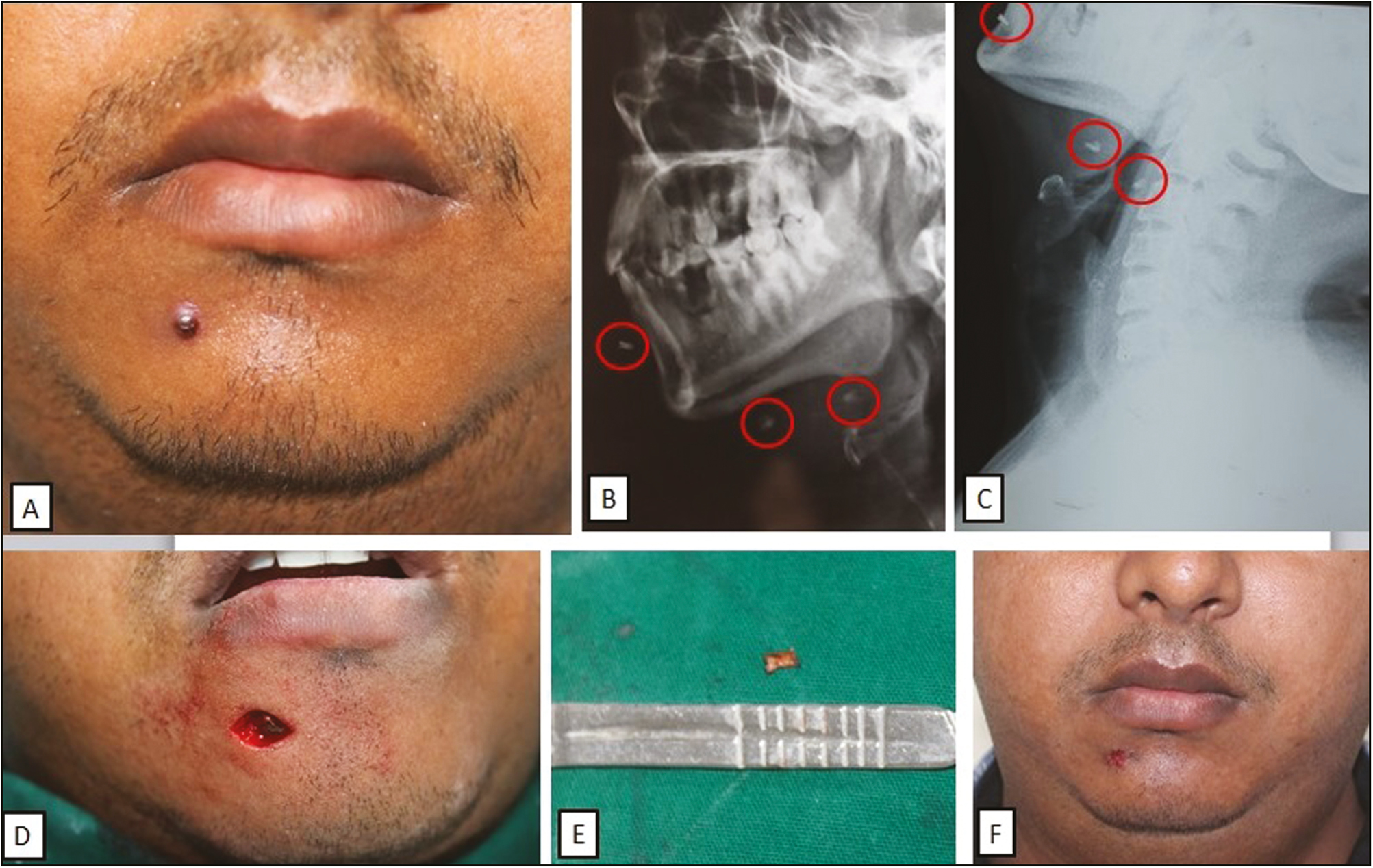
A 30-year-old male patient reported with a chief complaint of growth of small mass below the lower lip since 1 month. He had sustained minor facial injury due to a small blast while repairing an air conditioner 2 months back. On local examination, a small nodular growth measuring 0.5cm in diameter was present below the lower lip [Figure 2A]. No pus discharge was present. Lateral oblique X-ray view of the mandible and soft tissue neck X-ray revealed multiple (three) small radio-opacities, one at the anterior mandibular region and two in cervical region [Figure 2B, C]. Ultrasonography of the region was suggestive of collection measuring 1.3cm x 0.8cm and also revealed a blind-ending linear tract which extended from this collection to the soft tissue of the neck.

- (A) Preoperative view. (B) Lateral oblique X-ray confirming presence of multiple radio-opaque foreign objects. (C) Soft tissue neck X-ray confirming presence of multiple radio-opaque foreign objects. (D) Visible embedded foreign body through the existing wound. (E) Foreign body which turned out to be a splintered metallic particle. (F) One week follow-up
CASE 3
A 10-year-old female child reported to our clinic with alleged history of fall from a bicycle in the park. She had sustained injury over the left paranasal region. On local examination, contused lacerated wound was present over the left paranasal region [Figure 3A]. On palpation, presence of some foreign body was suspected.

- (A) Preoperative view. (B) Foreign body which turned out to be a wooden splinter. (C) Closure
Under local anesthesia, surgical access was made through the existing wound and once located precisely, the embedded foreign bodies were grasped with a hemostat and were retrieved out successfully in all three cases [Figures 1D, 2D]. The retrieved object turned out to be a glass particle, a splintered metallic particle, and a wooden splinter [Figures 1E, 2E, 3B]. The wound was closed in layers [Figures 1F, 3C]. The patients were prescribed routine antibiotics and analgesics. Injection Tetanus Toxoid was administered intramuscularly. Wound healing was satisfactory after 1 week in all three cases [Figures 1G, 2F]. In case 2, however, two foreign objects diagnosed in the cervical region were not retrieved.
DISCUSSION
Foreign objects can penetrate deep into soft and hard tissues through open wounds and lacerations which are sustained during trauma. If these foreign bodies are left undiagnosed in the tissues, they can result in serious complications within few days, months, or even years after the initial trauma.[4] In any non-healing wound resulting from penetrating injury that is showing continuous purulent discharge, having pain or developing a chronic draining sinus, the presence of a retained foreign body should be suspected.[5]
Plain radiographs, computed tomographic (CT) scan, ultrasonography, and magnetic resonance imaging (MRI) are helpful diagnostic tools to confirm the presence, location, size, and shape of foreign body.[2] Plain radiographs have detection success rate of 69–90% for metallic foreign bodies and 71–77% for glass cases; however, little or no information is available regarding the identification of organic material such as wood (0–15%).[3] Niamtu et al.[6] reported a case in which the styloid process simulated avulsed mandibular canine in a trauma patient on cervical spine radiograph, hence careful radiographic examination is mandatory to rule out normal anatomical structures.
Infection resulting from the retained wooden particle may lead to complications such as abscess and fistula formation. Ultrasound is a good diagnostic modality in detecting wooden foreign bodies in the maxillofacial region.[7] Metallic objects are radio-opaque and are mostly clearly visible on plain radiographs itself. It is advisable to get a CT scan done for precise location, anatomic proximation, and accurate diagnosis of these metallic objects. Use of MRI should be avoided in case metallic foreign body is suspected because MRI can mobilize metallic structures due to the magnetic field.[3]
When the impacted foreign body is superficially present and it is not lying near any major blood vessel, it can be removed under local anesthesia. The wound should be explored, foreign body should be removed, hemostasis should be achieved, followed by copious saline irrigation and suturing. It is advisable to prescribe antibiotic coverage as well as tetanus prophylaxis.[3]
In our cases, an early diagnosis was made on the basis of clinical examination and plain radiographs. In the first case, due to misdiagnosis at first instance, a glass piece was left which lead to infection. Later, it became clear that the glass piece was part of the spectacles which broke during the accident. In the second case, the foreign metallic particle was asymptomatic for 1 month. Later, it led to the development of nodular swelling. In the third case, a small wooden twig got impacted at the injury site. All three cases were treated under local anesthesia. All cases had uneventful recovery.
In the second case, two splintered foreign metallic particles were not retrieved due to close approximation to vital structures, surgical complexity of the cervical region, and asymptomatic feature [Figure 2B, C]. Inorganic foreign bodies, close approximation to vital structures like carotid artery, cervical spine, posterior orbit, and other structures, undue risk of iatrogenic injury, unknown precise location of foreign object, and absence of symptoms are certain contraindications for removal of embedded foreign objects in maxillofacial region.[8]
CONCLUSION
Early diagnosis and timely removal of impacted foreign bodies avoids functional, allergic, as well as infective complications.
Declaration of patient consent
The authors certify that they have obtained all appropriate patient consent forms. In the form the patient(s) has/have given his/her/their consent for his/her/their images and other clinical information to be reported in the journal. The patients understand that their names and initials will not be published and due efforts will be made to conceal their identity, but anonymity cannot be guaranteed.
Financial support and sponsorship
Nil.
Conflicts of interest
None.
Acknowledgement
None.
REFERENCES
- Management of foreign bodies in the maxillofacial region: Diagnostic modalities, treatment concepts with report of 2 cases. J Head Neck Physicians Surg. 2015;3:15-22.
- [Google Scholar]
- Unusual foreign bodies in the oral cavity: A report of three cases. Sch J Dent Sci. 2015;2:126-9.
- [Google Scholar]
- Impacted foreign bodies in the maxillofacial region-diagnosis and treatment. J Craniofac Surg. 2011;22:1404-8.
- [Google Scholar]
- Intraorbital bamboo foreign body in a chronic stage: Case report. Int J Oral Maxillofac Surg. 2000;29:428-9.
- [Google Scholar]
- Interesting case: Foreign body in the tongue. Br J Oral Maxillofac Surg. 2005;43:409.
- [Google Scholar]
- Styloid processes simulating avulsed tooth in a trauma patient. Oral Surg Oral Med Oral Pathol. 1984;58:240-1.
- [Google Scholar]
- Unusual foreign bodies in the orofacial soft tissue spaces: A report of three cases. Niger J Clin Pract. 2013;16:381-5.
- [Google Scholar]
- Traumatic foreign body into the face: Case report and literature review. Case Rep Dent. 2017;2017:3487386.
- [Google Scholar]


