Translate this page into:
Excision of Subungual Glomus Tumor by Subungual Approach: A Useful Yet Underutilized Technique
Address for correspondence: Dr. G. I. Nambi, Department of Plastic, Burns, Hand and Microsurgical Services, Kavin Medical Center, Perundurai Road, Erode 638011, Tamil Nadu. E-mail: nambi75@rediffmail.com
This is an open access journal, and articles are distributed under the terms of the Creative Commons Attribution-NonCommercial-ShareAlike 4.0 License, which allows others to remix, tweak, and build upon the work non-commercially, as long as appropriate credit is given and the new creations are licensed under the identical terms.
This article was originally published by Wolters Kluwer - Medknow and was migrated to Scientific Scholar after the change of Publisher.
Abstract
Abstract
Objective:
The aim of this study was to present an underutilized and underreported surgical technique in which the glomus tumors situated anywhere under the nail bed can be approached and removed with relative ease.
Materials and Methods:
Over 3 years, four cases of subungual glomus tumors, which were surgically managed with this technique were presented. The technical ease, complications, and aesthetic appearance of the nail were studied and presented. The limitation of this study was the lesser volume of cases.
Results:
This technique is easy to apply and gives aesthetically good results.
Keywords
Glomus tumor
lateral subperiosteal excision
subungual excision
subungual tumor
transungual excision
Introduction
Subungual glomus tumors are rare neoplasms of the digits. Definitive management lies in the complete removal of the tumor and curettage of the cavity. Two common types of excisions are performed depending on the location of the tumor under the nail bed. For centrally located lesions, the transungual approach is adopted and for lateral or peripherally located lesions, the lateral periosteal approach is adopted.[123456] Another type of excision by way of subungual approach[7] in which lesions located anywhere under the nail bed can be approached with relative ease and gives aesthetically good results is presented in this article.
Materials and Methods
Four (three males and one female) cases of subungual tumors that were operated between March 2014 to May 2017, were retrospectively studied. In all the cases, continuous and excruciating pain was the predominant symptom. The pain aggravated when the hand was put to use, on dependency, and also when coming in contact with cold objects. Sleep disturbance in the night because of pain was noted in two cases. The dominant hand was involved in two cases and the nondominant hand was involved in the other two. The thumb was the digit affected in the nondominant hands, whereas thumb and index finger were involved in the dominant hands. In all the cases, the clinical diagnosis was confirmed with radiograph and magnetic resonance imaging (MRI) [Figures 1 and 2]. The tumor was centrally located in three cases and on the ulnar side of the nail bed in one case. All the cases were operated using the following technique.

- Plain X-ray of the digit shows a clear halo in the bone due to erosion
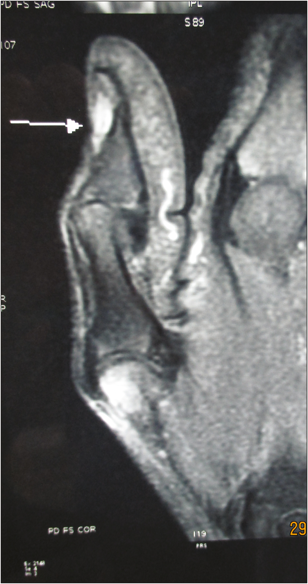
- MRI image of the tumor showing hyperintense signal in T1-weighted sequence with increased contrast uptake
Technique: In all the cases, digital block and digital tourniquet were used. The nail was separated from the nail bed and the dorsal skin by passing a mosquito artery forceps and was uprooted intact and preserved. The nail bed was raised as a proximally based flap along with the underlying periosteum in a distal-to-proximal direction, exposing the underlying tumor [Figure 3]. The tumor was removed in total, followed by curettage and washing of the cavity [Figure 4]. The nail bed, which was elevated as a proximally based flap, was replaced back and secured in the distal end with one or two absorbable sutures (4-0 rapid Vicryl with cutting needle was used in all of our cases). The uprooted intact nail was placed back in its position and was secured with a half-buried horizontal mattress suture passing via skin, germinal matrix, and then back to the skin [Figure 5]. One simple suture was applied in the middle of the distal end (all using the same 4-0 rapid Vicryl). The digital tourniquet was released only after this and then the dressings were carried out.
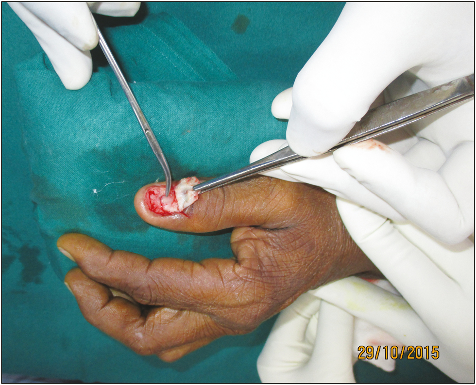
- After uprooting the nail, the nail matrix along with the underlying periosteum is raised as proximally based flap exposing the underlying tumor. The tumor is being pointed with a curved artery forceps and the flap is being held in thumb forceps
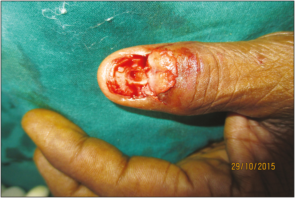
- Cavity after curettage
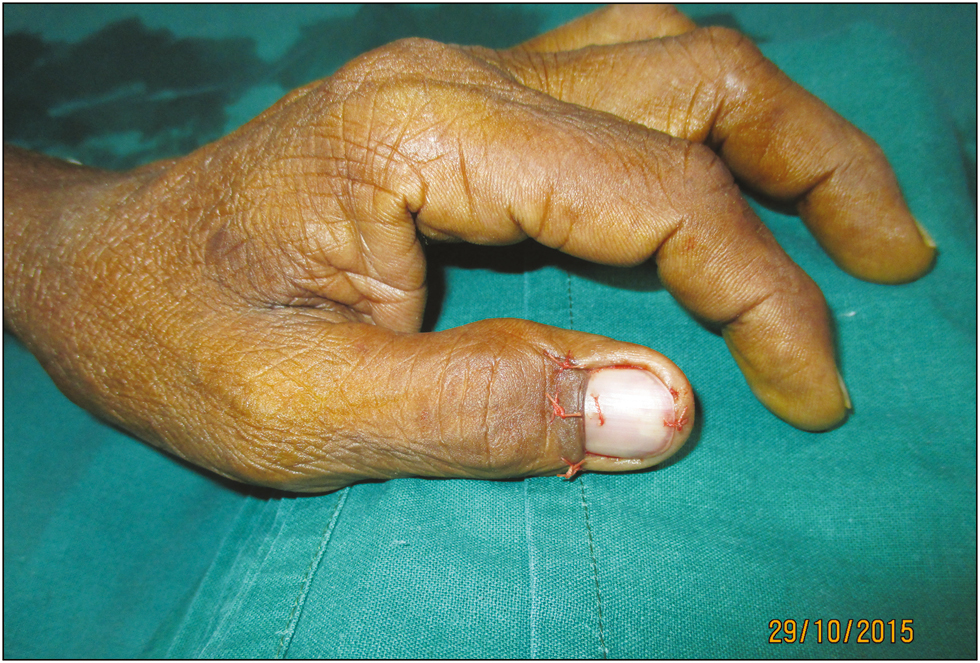
- Uprooted nail is replaced back and is held with sutures
Results
The average duration of the procedure was 30min. Complete pain relief was the most significant factor in the post-op period, and the happiest were the ones who had sleepless nights due to pain. The follow-up period was from 3 months to 1 year, and no episodes of post-op infection, nail deformity, nail loss, or recurrence of symptoms were reported [Figure 6].
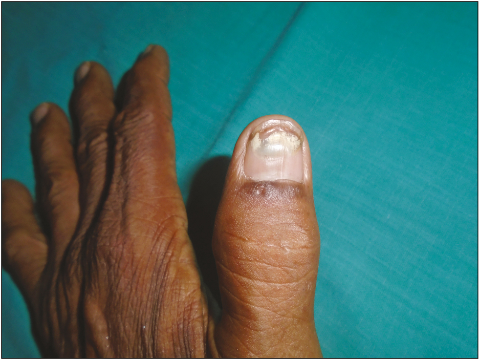
- Six months postoperative follow-up picture
Discussion
Subungual glomus tumors are rare benign neoplasms present under the nail bed and constitute 1%–5% of hand tumors. They arise from glomus bodies, which are thermoregulatory apparatuses. Though they can occur anywhere in the body, 50% of them are located in the subungual region of the digits. Depending on their location in the nail bed, subungual tumors can be called central or peripheral. Excruciating continuous pain, which aggravated on exposure to colder temperature, is the most common presentation. Bluish discoloration of a focal area of nail, warmth, tenderness to palpate, and increased sensitivity to cold touch are common clinical findings. MRI is the gold standard radiological investigation to confirm the clinical diagnosis, and if the underlying bone is eroded by the tumor, plain radiogram shows a clear halo and is sufficient to clinch the diagnosis.[1234567]
Surgical excision is the definitive management and depending on the location of the subungual glomus tumor, two common techniques are being followed. For centrally located lesions, a transungual approach is used in which after removing the nail, a vertical incision is made over the nail bed overlying the tumor and the tumor is exposed. For peripheral or laterally located lesions, a lateral subperiosteal approach is used in which after removing the nail, the incision is made along the lateral border of the nail bed and is lifted up to expose the tumor. After the tumor is excised and the cavity is curetted, washed, and the wound is closed in layers, the overlying nail can be retained or discarded. The advantage of preserving the overlying nail is that there is less postoperative pain and it protects the underlying nail bed from exposure and trauma.
In the subungual approach presented in this article, the nail bed is raised with the underlying periosteum as a proximally based flap in a distal-to-proximal direction. The advantages of this technique are as follows:
The subungual approach allows for the complete exposure of the underlying bone and the glomus tumors, which are located anywhere under the nail bed, can be approached with ease.
By lifting the nail bed as a proximally based flap, there is adequate space provided to the surgeon to operate in a very limited operating field. With adequate exposure, chances of recurrence because of inadequate curettage are avoided.
As the entire nail bed is raised as flap and inset back in its place after the procedure, the later nail surface is smooth without serrations.
The replaced nail protects the underlying nail bed and provides a good scaffold for the future nail.
To conclude, though the subungual approach to glomus tumor excision was described long ago,[7] hardly any reports were noted in the literature. In all the cases reported in this study, this approach was used with ease and with good results but with short case volume, more inputs are needed in terms of long-term outcome and comparative studies with other techniques.
Declaration of patient consent
The authors certify that they have obtained all appropriate patient consent forms. In the form the patient(s) has/have given his/her/their consent for his/her/their images and other clinical information to be reported in the journal. The patients understand that their names and initials will not be published and due efforts will be made to conceal their identity, but anonymity cannot be guaranteed.
Financial support and sponsorship
Nil.
Conflicts of interest
There are no conflicts of interest.
References
- Subungual glomus tumors of the hand: treated by transungual excision. Indian J Orthop. 2015;49:403-7.
- [Google Scholar]
- Subungual glomus tumours: diagnosis and microsurgical excision through a lateral subperiosteal approach. J Plast Reconstr Aesthet Surg. 2014;67:373-6.
- [Google Scholar]
- Nail-preserving excision for subungual glomus tumour of the hand. J Plast Surg Hand Surg. 2014;48:201-4.
- [Google Scholar]
- Which approach is best for subungual glomus tumors? Transungual with microsurgical dissection of the nail bed or periungual? Chir Main. 2015;34:39-43.
- [Google Scholar]
- Subungual glomus tumors: surgical approach and outcome based on tumor location. Dermatol Surg. 2013;39:1017-22.
- [Google Scholar]
- The perionychium. In: Green DP, Hotchkisss RS, Pederson WC, Wolfe SW, eds. Green’s operative hand surgery (5th ed). Philadelphia, PA: Elsevier; 2005. p. :389-416.
- [Google Scholar]


