Translate this page into:
Basal Cell Carcinoma Camouflaged by Rhinophyma
Address for correspondence: Adelina Costin, Department of Dermatology and Venereology, Hospital Garcia de Orta, Av. Torrado da Silva, 2805-267 Almada, Portugal. E-mail: adelinacostin@gmail.com
This is an open access journal, and articles are distributed under the terms of the Creative Commons Attribution-NonCommercial-ShareAlike 4.0 License, which allows others to remix, tweak, and build upon the work non-commercially, as long as appropriate credit is given and the new creations are licensed under the identical terms.
This article was originally published by Wolters Kluwer - Medknow and was migrated to Scientific Scholar after the change of Publisher.
Dear Editor,
Rhinophyma is a rare and progressively disfiguring condition, characterized by sebaceous hyperplasia and fibrous tissue proliferation.[1] In its extreme form, it may cause nasal airway obstruction and hide malignancies, in addition to the cosmetic and emotional concerns.
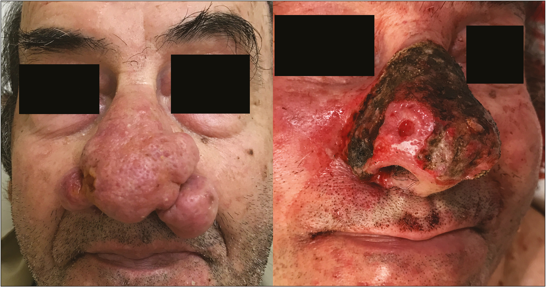
An 87-year-old man, with a long-standing history of rosacea, presented with exuberant rhinophyma on the tip of the nose. A closer look disclosed a small crusted papule on the right ala [Figure 1]. The clinical differential diagnosis included phimatous nodule, basal cell carcinoma, or adnexal tumors.

- Exuberant rhinophyma with an eroded nodule on the right ala before and after electro-rhinosculpture
To achieve negative margins and plan the reconstructive approach, we first performed an electro-rhinosculpture. After anesthesia with injection of 2% lidocaine with 1:100,000 epinephrine, the phimatous nodules were excised with a loop tip of electrosurgical device in cutting mode, thus allowing to remove the excess tissue in smooth planes while providing simultaneous hemostasis. Mechanical dermabrasion followed using the cautery tip polisher in order to soften the nasal contours into the desired appearance [Figure 1]. Additional hemostasis was performed with electrocoagulation. We then performed a dressing with metronidazole cream and Surgicel.
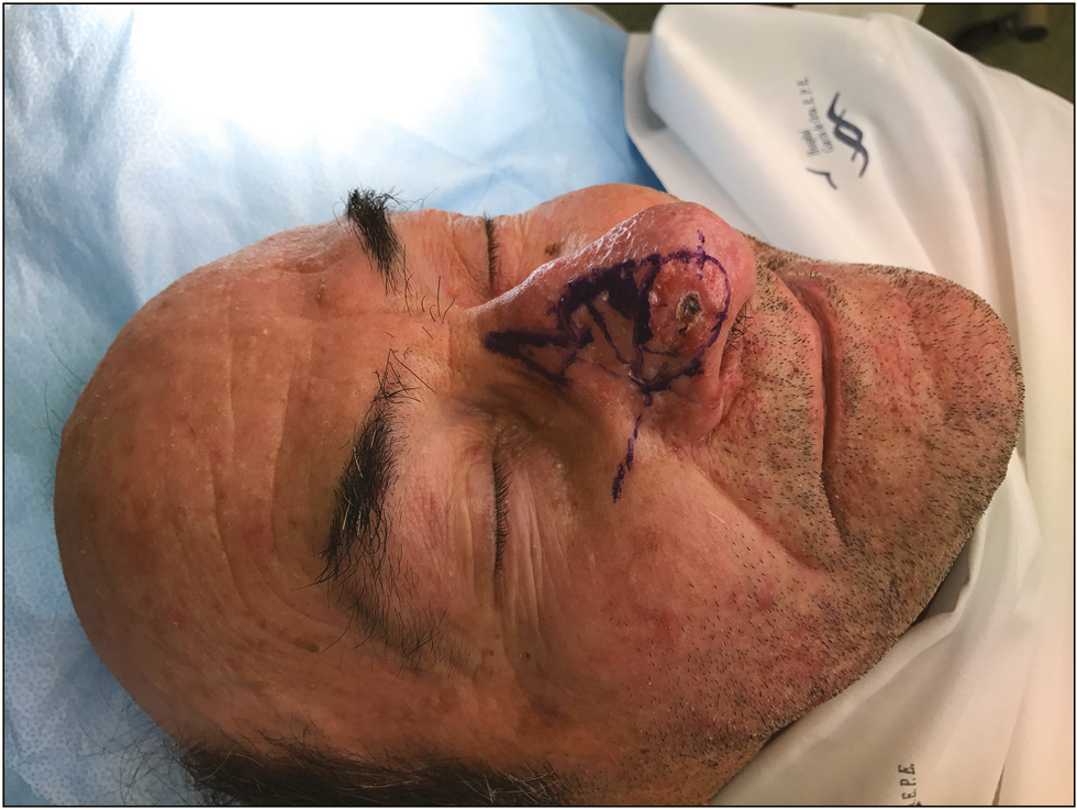
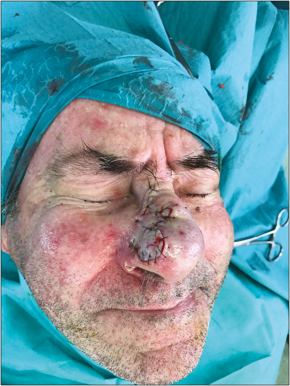
In a deferred time, we performed tumor excision and reconstructed the surgical defect using a bilobed flap [Figures 2 and 3].

- Reconstruction with a bilobed flap after excision of basal cell carcinoma

- Reconstruction with a bilobed flap after excision of basal cell carcinoma
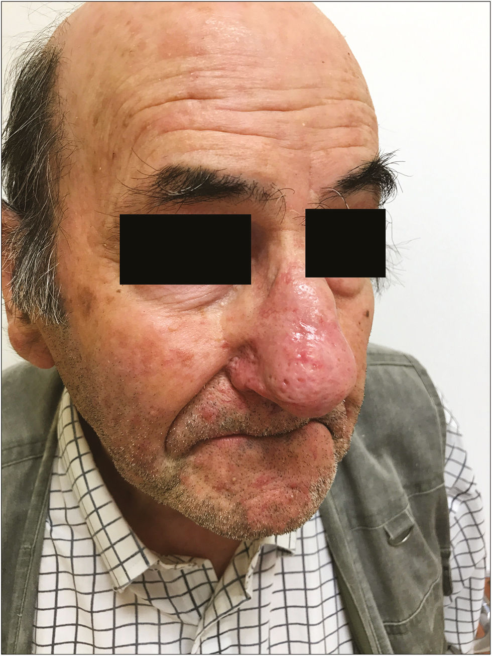
At 4-week follow-up, the patient had normal nasal contour, with excellent functional and cosmetic result [Figure 4]. There was no significant scarring on our patient. To avoid scarring, excision should not be below the depth of the pilosebaceous unit, which would result in atrophy rather than porous nasal skin.

- Four weeks after treatment
To the best of our knowledge, there are a few reports of an association between rhinophyma and basal cell carcinoma.[2] It is important to acknowledge this as the surgeon should be alert to the presence of malignancy in atypical or rapidly enlarging rhinophyma in order to guarantee the best treatment approach.
Declaration of patient consent
The authors certify that they have obtained all appropriate patient consent forms. In the form the patient(s) has/have given his/her/their consent for his/her/their images and other clinical information to be reported in the journal. The patients understand that their names and initials will not be published and due efforts will be made to conceal their identity, but anonymity cannot be guaranteed.
Financial support and sponsorship
Nil.
Conflicts of interest
There are no conflicts of interest.
REFERENCES
- Rosacea. Part I: Introduction, categorization, histology, pathogenesis, and risk factors. J Am Acad Dermatol. 2015;72:749-58. quiz 759-60
- [Google Scholar]
- Basal cell carcinoma mimicking rhinophyma: Case report and literature review. Arch Dermatol. 1988;124:1077-9.
- [Google Scholar]


