Translate this page into:
Healing of a Large Wound Defect Post Debridement, with PRF Therapy and High Dose Oral Vitamin C, in a Patient of Severe Irritant Contact Dermatitis Due to Slaked Lime: A Case Report
Address for correspondence: Mr. Shashank Bansod, Hi-Tech Skin Clinic and Hair Transplant Centre, 3rd Floor, Vijay Bhawan, Above Jain Medicals, Opposite Lokmat Bhawan, Wardha Road, Nagpur 440010, Maharashtra, India. E-mail: hitech.skin@yahoo.com
This is an open access journal, and articles are distributed under the terms of the Creative Commons Attribution-NonCommercial-ShareAlike 4.0 License, which allows others to remix, tweak, and build upon the work non-commercially, as long as appropriate credit is given and the new creations are licensed under the identical terms.
This article was originally published by Wolters Kluwer - Medknow and was migrated to Scientific Scholar after the change of Publisher.
Abstract
Abstract
Platelet-rich blood concentrates have been used to accelerate healing process in wounds and in bones since many decades worldwide. Platelet-rich fibrin (PRF) is a relatively new and established therapy, utilizing platelets and leucocytes trapped in fibrin matrix, for the treatment of non-healing ulcers and wounds. Many large series are available in this subject to prove its efficacy. Our patient, a known case of eczema, had applied slaked lime (calcium hydroxide) over an eczematous lesion on right leg and surrounding area, after which he developed deep wound with extensive erythema and blisters initially, which healed with necrosis due to patient’s neglect, in about 2 weeks. On presentation to us, the lesion had undergone necrosis and hence decision to debride the lesion was taken. After debridement, a large defect was created, which we tried treating conservatively using PRF therapy primarily, followed by pressure dressing. High dose vitamin C was given orally. The patient required antibiotics intermittently. The patient responded well to this protocol and the wound defect was closed within a few weeks.
Keywords
Debridement
PRF therapy
vitamin C
wound healing
INTRODUCTION
Slaked lime or calcium hydroxide, a commonly eaten substance in Indian household, along with betel-nut leaf, is a known cause of contact irritant dermatitis and can cause massive and deep skin burns.[1]
If the lesion undergoes necrosis, as in our case, debridement followed by skin grafting is a commonly followed treatment.[2]
Platelet-rich fibrin (PRF) is an established therapy for the treatment of non-healing wounds and ulcers, which involves slow release of cytokines trapped in fibrin mesh, helping in different aspects of wound healing.[3]
Vitamin C has a proven role in wound healing; it helps in collagen synthesis and remodeling.
Conservative management of large and deep wounds after debridement, using PRF therapy and high dose oral vitamin C, was attempted considering their established role in wound healing.
CASE REPORT
A 52-year-old man presented with an asymptomatic, large, crusted lesion over right leg few inches below the knee [Figure 1]. On eliciting history, patient was a known case of eczema and had applied slaked lime (calcium hydroxide) liberally over eczematous lesion and surrounding area of foot, after which he developed extensive redness and blistering over that area, which reduced spontaneously, leaving behind large adherent crust over and nearby the area of application. Personal history, past medical history, and drug history were not relevant
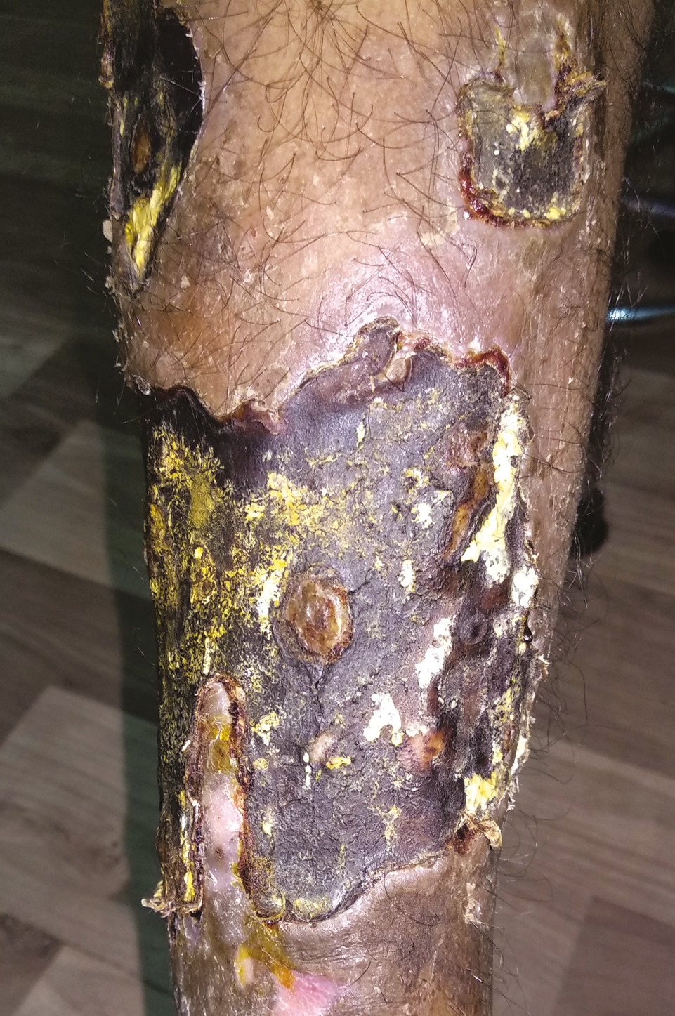
- At presentation. Large lesion with overlying crust
On physical examination, patient had one large lesion of approx. 17 cm × 8 cm and one small lesion of size approx. 8 cm × 6 cm; both lesions were present over the right leg extending from below the knee till the shin area.
Each lesion was firm-to-hard in consistency with a thick crust covering it. After removing the crust from a small part, it revealed a deep necrotic area underneath. We decided to debride the lesion and try to let the wound defect heal conservatively, using PRF therapy initially.
Mechanical debridement was done to remove necrotic tissue under spinal anesthesia, under all aseptic precautions [Figure 2], and a specimen was sent for histopathological analysis, whose findings were consistent with contact dermatitis.
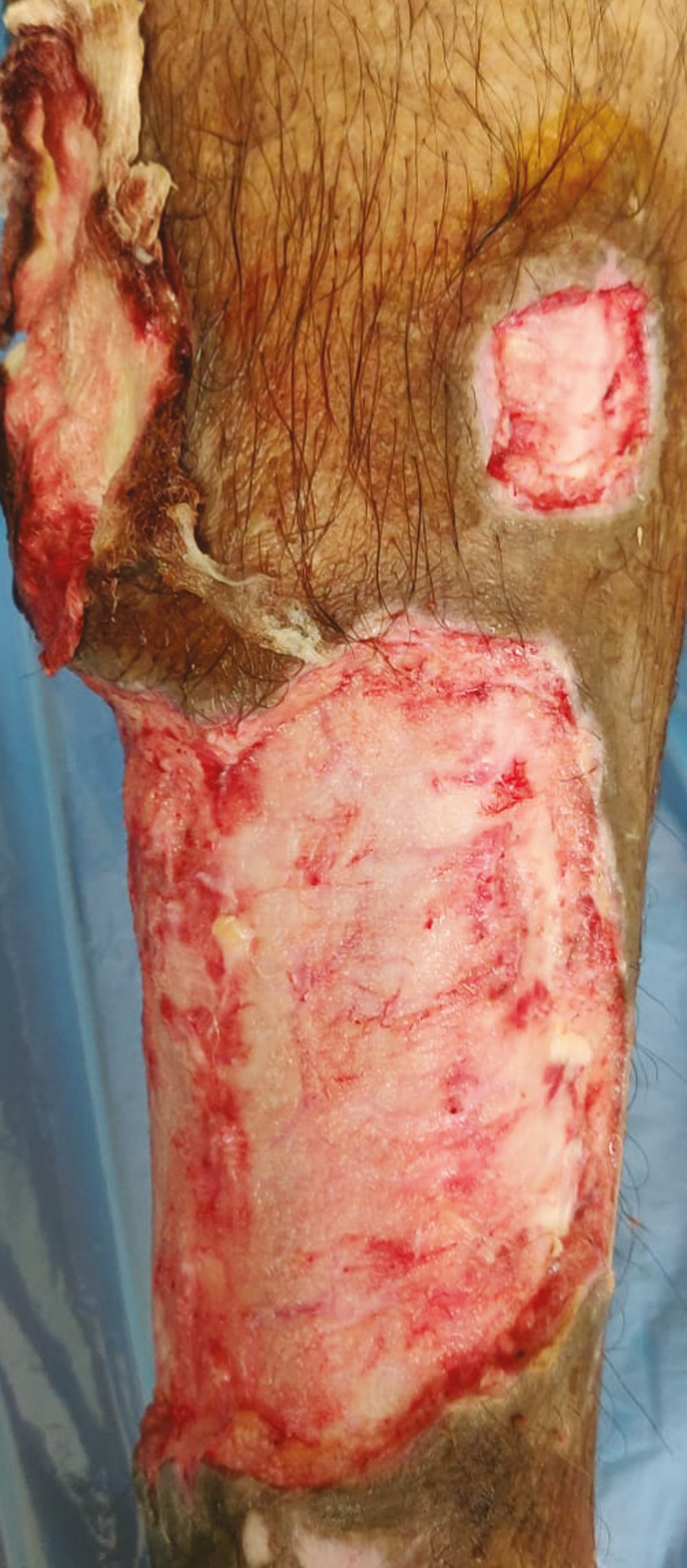
- Immediately after debridement. A large wound defect is seen
The wound defect thus created was dressed with a three-layered wet dressing.
Magnesium sulfate-impregnated gauze pieces were placed over the wound, followed by a thick absorbent cotton pad, which was held in place firmly by a gauze wrap. The dressing was changed every 3 days, till 1 week, when the oozing from the wound reduced and thereafter the first session of PRF was planned [Figure 3].
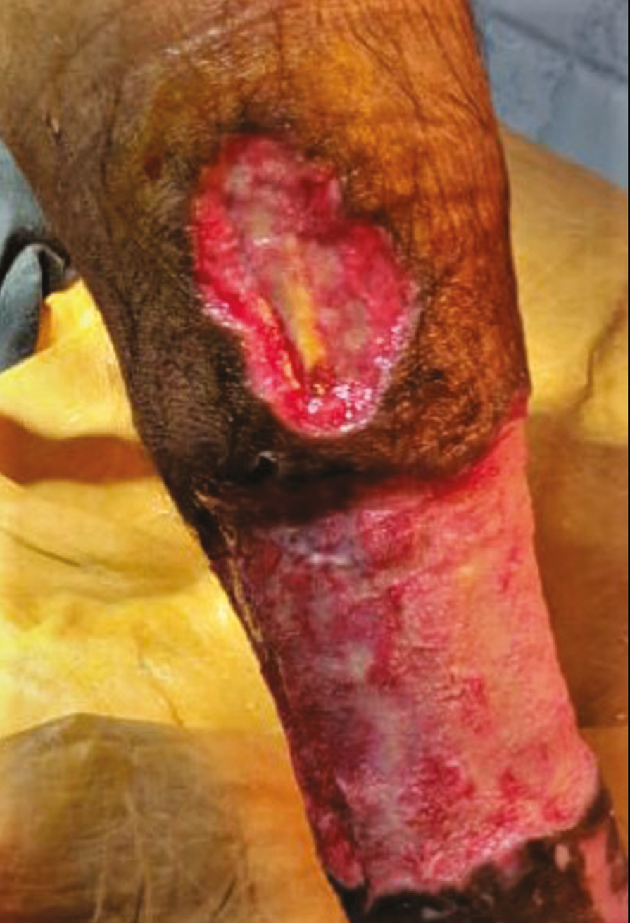
- One week after debridement. Stable wound without oozing
PRF used was L-PRF or leucocyte-rich PRF and was prepared using modified Choukran’s protocol.[4] Since the size of the wound defect was very large, we divided it into three sections for our convenience.
PRF was done on one wound section at a time initially, after which the size of the wound decreased.
Using scalp vein catheter and syringe, 30 mL of blood was drawn from the patient under all aseptic precautions, which was collected in two 15 mL conical glass tubes. After balancing the tubes of centrifuge (Remi R4C by Remi Labs), the tubes were rotated at 3000 rpm for 10 min.
At the end of centrifugation, three layers were seen in the tube: upper plasma layer, PRF clot as a middle layer, and lower layer of RBCs.
The PRF clot was obtained immediately after centrifugation, which, after being taken out from the tube carefully using a pair of forceps, was pressed between two gauze pieces firmly, so as to form a membrane.
The membrane was then cut in small pieces and each piece was placed over the wound section randomly. The wound was closed with a triple-layered dressing as mentioned earlier.
The PRF therapy was repeated on the second wound section after 7 days and on the third wound section 7 days after that. The depth and size of the wound started reducing at the end of first week.
At the end of 3 weeks, the wound defect was visibly reduced and filled up with granulation tissue.
Two more PRF sessions were done within a gap of 10 days over the whole defect, starting 7 days after the previous session.
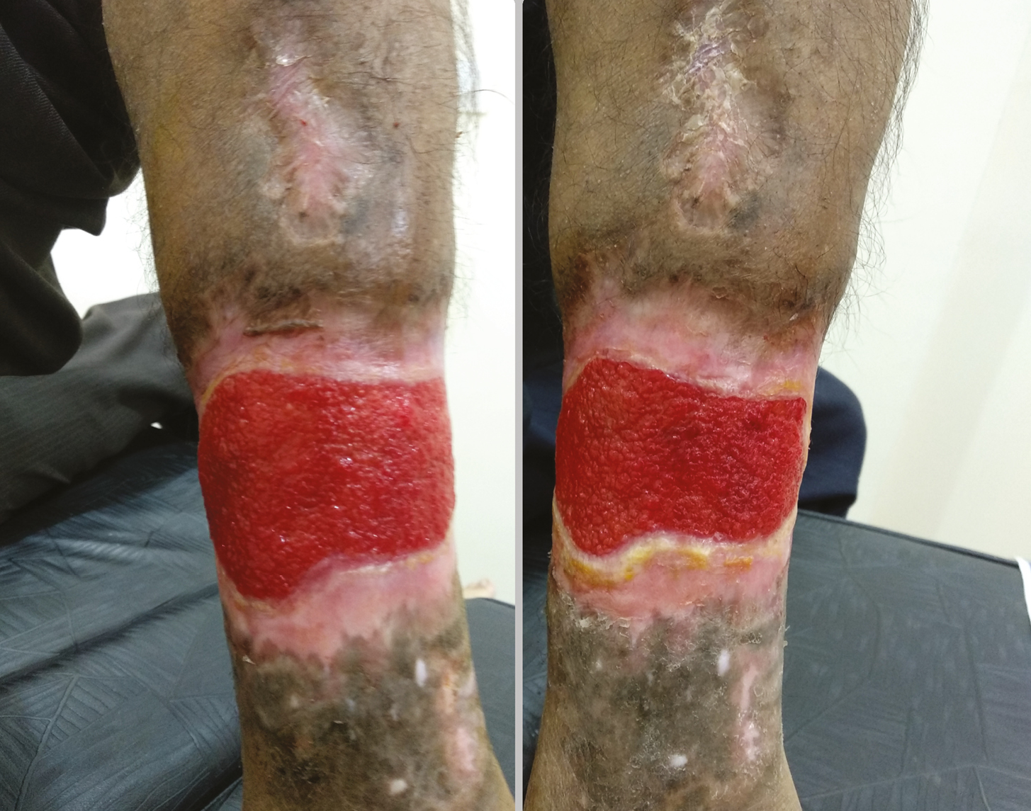
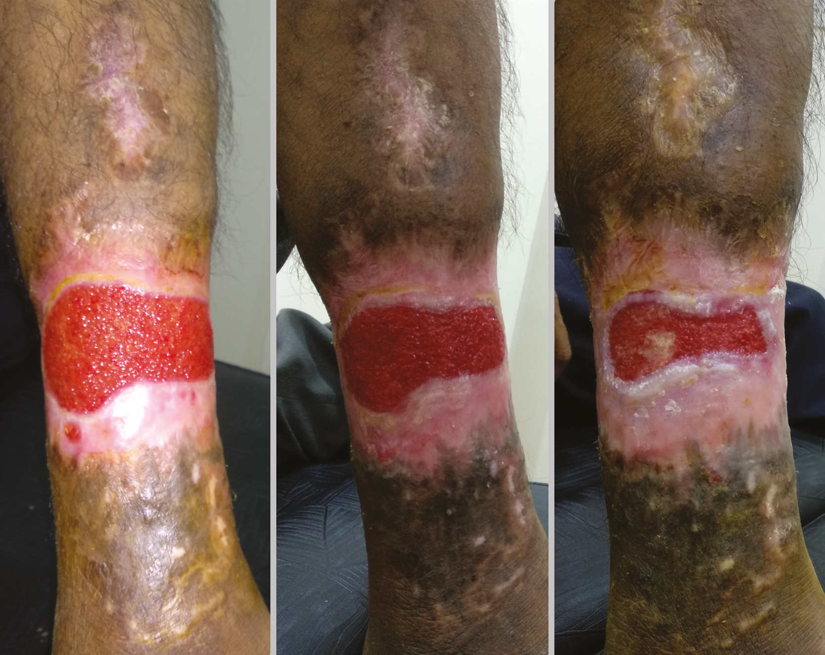
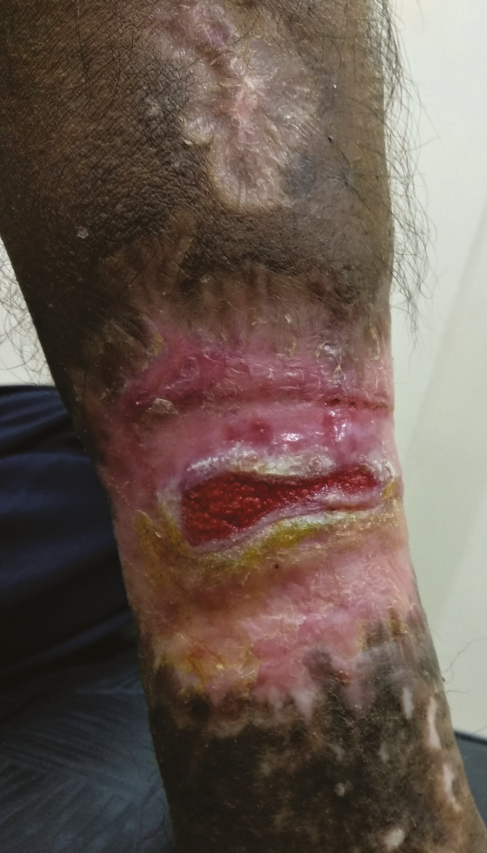
A total of five sessions of PRF therapy were done. Thereafter, only wound care comprising of cleaning of wound with hydrogen peroxide solution and rubbing out excess granulation tissue with gauze pieces, followed by a three-layered dressing was performed every 7 days. The wound was fully filled up in around 10 weeks’ time [Figures 4–8].
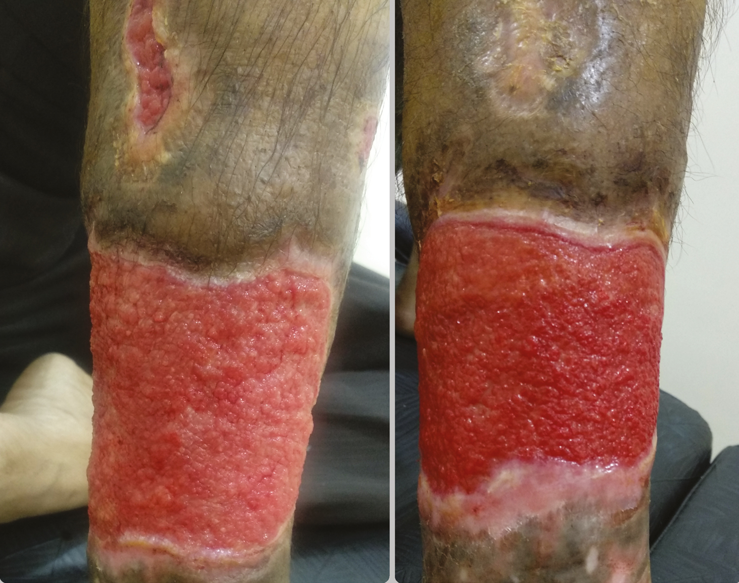
- Weeks 2 and 3. Progression of healing
- Weeks 4 and 5 after therapy. Visible reduction of defect
- Weeks 6–8: progressive healing
- Week 9, wound almost healed
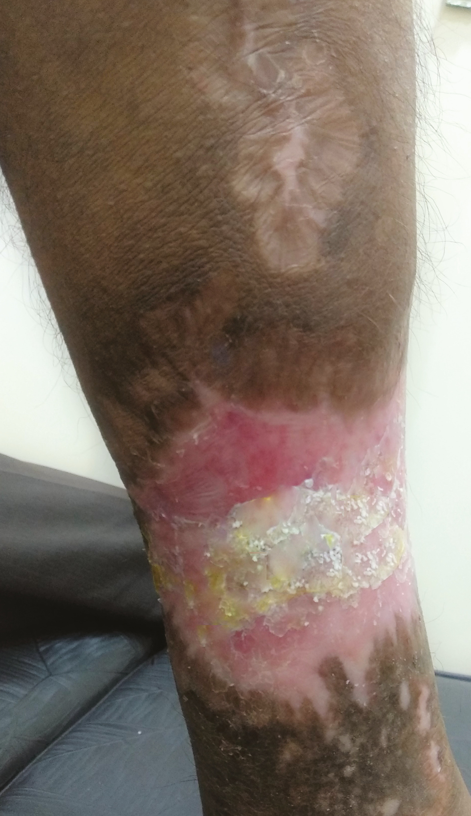
- Week 10, defect closed
The patient also received 1000 mg of oral vitamin C daily, from day 1 of treatment till the end. Intermittently, oral antibiotics and analgesics were also given as required.
RESULT
Post debridement large wound defect was successfully closed conservatively using PRF therapy and oral vitamin C, with sustained results.
DISCUSSION
Calcium carbonate or slaked lime is a known cause of contact irritant dermatitis. Few minutes to hours of application causes erythema and blistering, especially if applied to area with breach of continuity of skin due to increased absorption of irritant.[5] If not treated promptly, the lesion may heal with necrosis.
Conservative treatment to surgical options is available depending on wound condition. If necrosis sets in, mechanical debridement followed by skin grafting is done.
We tried to use PRF as a rescue therapy for quicker wound healing conservatively. PRF, a fully autologous product, was first used by Choukran et al. in France[6] for the treatment of non-healing wounds.
There are two traditional types of PRF, P-PRF in which the leucocytes are absent. It is tedious to make and not cost-effective. Another type is L-PRF or leucocyte-rich PRF, which is easy to prepare and very cost-effective. Choukran et al. added A-PRF or the advanced PRF to the mentioned types. This, he claimed, was superior in terms of cell distribution and healing properties. We have used L-PRF for facilitating wound healing in the mentioned case.
The proposed mechanisms for all types of PRF more or less include slow polymerization from fibrinogen to fibrin, within which the cytokines released by degranulated platelets are trapped and slowly released over a period of time, which helps in wound healing.[7]
Platelet-derived growth factor, vasculo-endothelial growth factor, fibroblast growth factor, and thrombospondin are some of the important cytokines released by platelets.[8]
PRF has been previously used in the healing of complicated necrotic-free graft in dentistry, similar scenario as in our case, but much less the wound defect.[9]
Vitamin C helps in all phases of wound healing right from inflammatory phase where it helps in neutrophil apoptosis to proliferative phase where it helps in collagen synthesis, maturation, and degradation.[10]
CONCLUSION
The article reiterates the role of platelet derivatives for extensive non-healing wound with good results. PRF therapy and oral vitamin C can be a good option in conservative management of very large wound defects.
Declaration of patient consent
The authors certify that they have obtained all appropriate patient consent forms. In the form, the patient(s) has/have given his/her/their consent for his/her/their images and other clinical information to be reported in the journal. The patients understand that their names and initials will not be published and due efforts will be made to conceal their identity, but anonymity cannot be guaranteed.
Financial support and sponsorship
Nil.
Conflicts of interest
There are no conflicts of interest.
REFERENCES
- Contact dermatitis and drug eruptions. Andrews’ diseases of the skin e-book: Clinical dermatology, Chapter 6, p.. :92.
- [Google Scholar]
- Burn debridement, grafting, and reconstruction. In: Stat Pearls [Internet]. Treasure Island, FL: Stat Pearls Publishing; 2020. [Updated 2020 Feb 12]
- [Google Scholar]
- Platelet-rich fibrin and its emerging therapeutic benefits for musculoskeletal injury treatment. Medicina (Kaunas). 2019;55:141. 10.3390/medicina55050141
- [Google Scholar]
- A proposed protocol for the standardized preparation of PRF membranes for clinical use. Biologicals. 2012;40:323-9. doi: 10.1016/j.biologicals.2012.07.004. Epub 2012 Jul 28
- [Google Scholar]
- Burn wound: How it differs from other wounds. Indian J Plast Surg. 2012;45:364-73. doi: 10.4103/0970-0358.101319
- [Google Scholar]
- Une opportunite′ en paro-implantologie: le PRF. Implantodontie. 2001;42:55-62 (in French).
- [Google Scholar]
- Platelet-rich fibrin application in dentistry: A literature review. Int J Clin Exp Med. 2015;8:7922-9.
- [Google Scholar]
- Platelet-rich concentrates differentially release growth factors and induce cell migration in vitro. Clin Orthop Relat Res. 2015;473:1635-43. 10.1007/s11999-015-4192-2
- [Google Scholar]
- Role of platelet-rich-fibrin in enhancing palatal wound healing after free graft. Contemp Clin Dent. 2012;3(Suppl 2):S240-3. 10.4103/0976-237X.101105
- [Google Scholar]
- Vitamin C: A wound healing perspective. Br J Commun Nurs (Suppl):S6-11. 10.12968/bjcn.2013.18. sup12.s6
- [Google Scholar]


