
Translate this page into:
Update in the Uses of Chalazion Clamp in Dermatological Procedure Revisited
Address for correspondence: Dr. Shreya K, All India Institute of Medical Sciences (AIIMS), Habibganj, Saketnagar, Bhopal 562020, Madhya Pradesh, India. E-mail: shreyakgowda@gmail.com
This is an open access journal, and articles are distributed under the terms of the Creative Commons Attribution-NonCommercial-ShareAlike 4.0 License, which allows others to remix, tweak, and build upon the work non-commercially, as long as appropriate credit is given and the new creations are licensed under the identical terms.
This article was originally published by Wolters Kluwer - Medknow and was migrated to Scientific Scholar after the change of Publisher.
Abstract
Abstract
Chalazion clamp has a distal tip with a flat oval plate; other side has a ring-like structure and forceps-like handle. Handle has a thumbscrew, which can be tightened and released so as to achieve the required pressure. Available in various sizes. Used mainly in ophthalmological procedures primarily. Used in dermatology by modifying its technique and creating an adequate hemostasis in various diagnostic and interventional procedures. In the previous article chalazion clamp was used for preparing SSS for ear lobule, so this article revisited the uses of chalazion clamp in dermatology and extended use in the preparation of adequate blanching while preparing SSS from the peripheral tissue site.
Keywords
Chalazion clamp
dermatology
SSS

INTRODUCTION
Slit Skin Smear from ear lobule and peripheral site from most infiltrated plaque is performed as routine OPD procedure, adequate smear can be obtained by pinching the ear lobule and pinching skin from the plaque which requires skill and manual power. Adequate blanching can be achieved by clamping the ear lobule and skin with chalazion clamp.
Chalazion clamps are either disposable or reusable. Available in three sizes, small (ring size 11 mm × 17 mm), medium (ring size 12 mm × 23mm), and large (ring size 17 mm × 28 mm).[1]
Chalazion clamp has a distal tip with flat oval plate; other side has a ring-like structure and forceps-like handle. Handle has a thumbscrew, which can be tightened and released so as to achieve the required pressure.
In the previous article chalazion clamp was used for preparing SSS for ear lobule, so in this article revisited the uses of chalazion clamp in dermatology and extended use in the preparation of adequate blanching while preparing SSS from peripheral tissue site.
Advantages of chalazion clamp over routine SSS preparation
Minimizes the bleeding and hence adequate smear is obtained.
Provides a firm immobile surface so that smear can be taken easily.
Achieve firm grip.
Screw clamp allows variability in the pressure applied compared to artery forceps
Uses of chalazion clamp
Opthalmology: Chalazion surgery
Dermatology
In our case we used in preparing SSS and achieved an adequate smear by using reusable chalazion clamp.
As reported previously sponge holding forceps and artery forceps does not provide required pressure may result in tissue crushing and excessive pain, so modified in our case with chalazion clamp.
Procedure
After getting informed consent from the patient, Chalazion clamp (as per Figure 1) applied to ear lobule (Figure 2) is tightened until it blanches, similarly done on other areas (Figure 3) from where smear is taken. The SSS is taken using surgical blade no 23. A 5 mm length, 2 mm depth incision given. Turn the blade 90°, scrape out fragments of tissue and fluid on glass slide.[5] A smear of 8–10 mm is prepared on glass slides. Chalazion clamp is released, skin cut is sealed with a small piece of cotton wool dipped in tincture of benzoin.
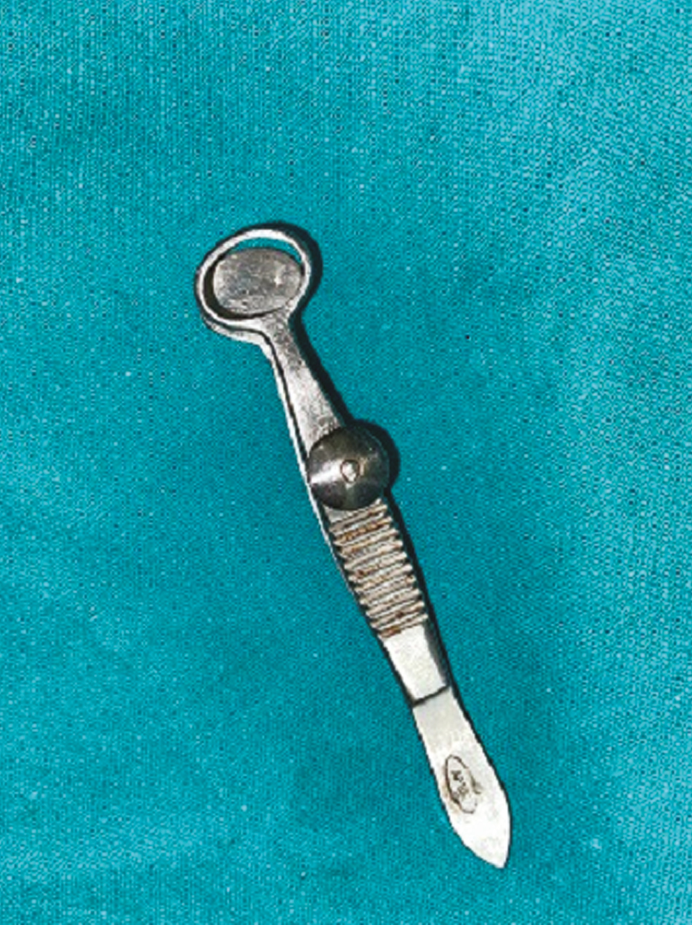
- Chalazion clamp small size
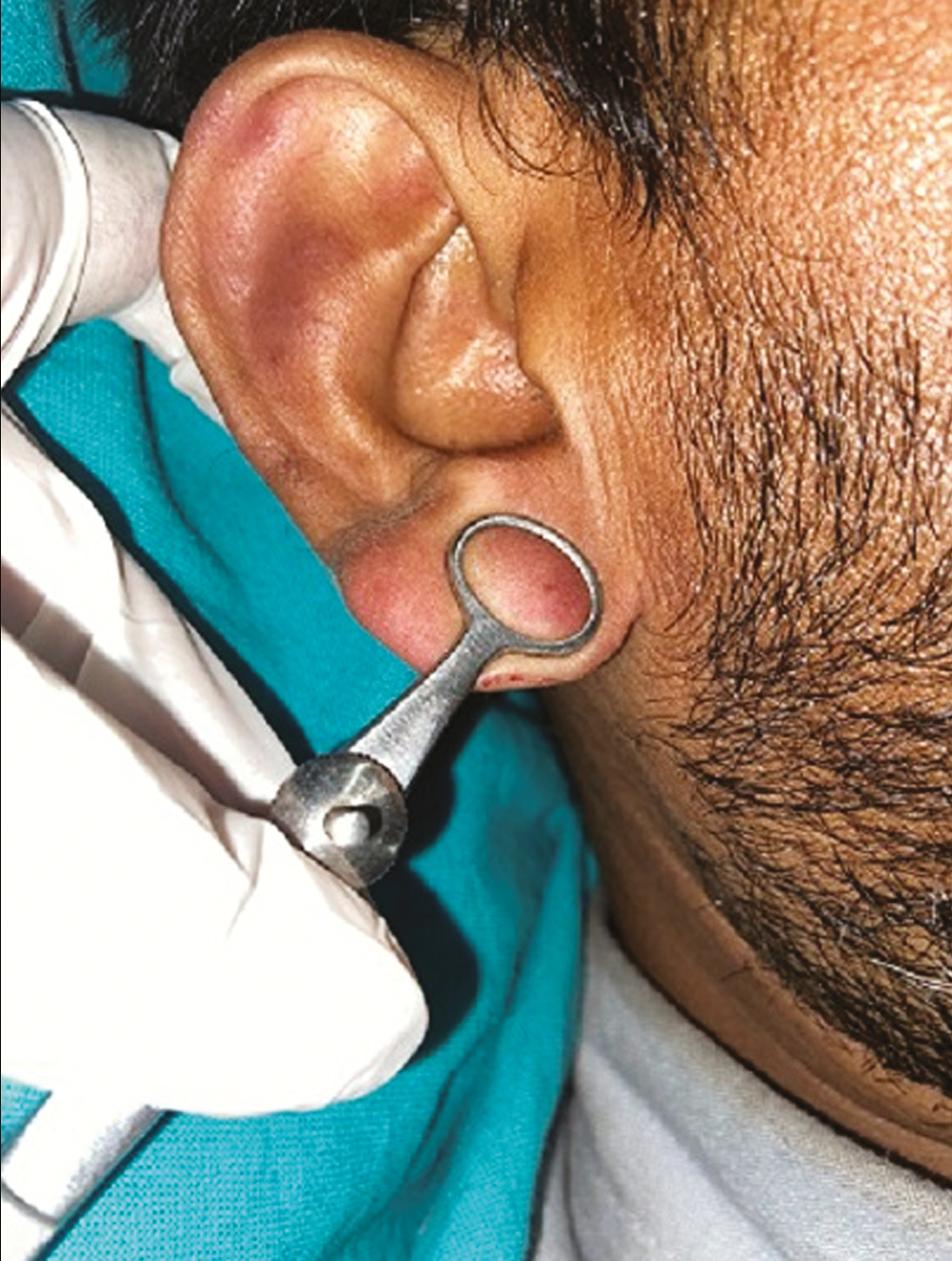
- Use of chalazion clamp in slit skin smear sampling in ear lobule
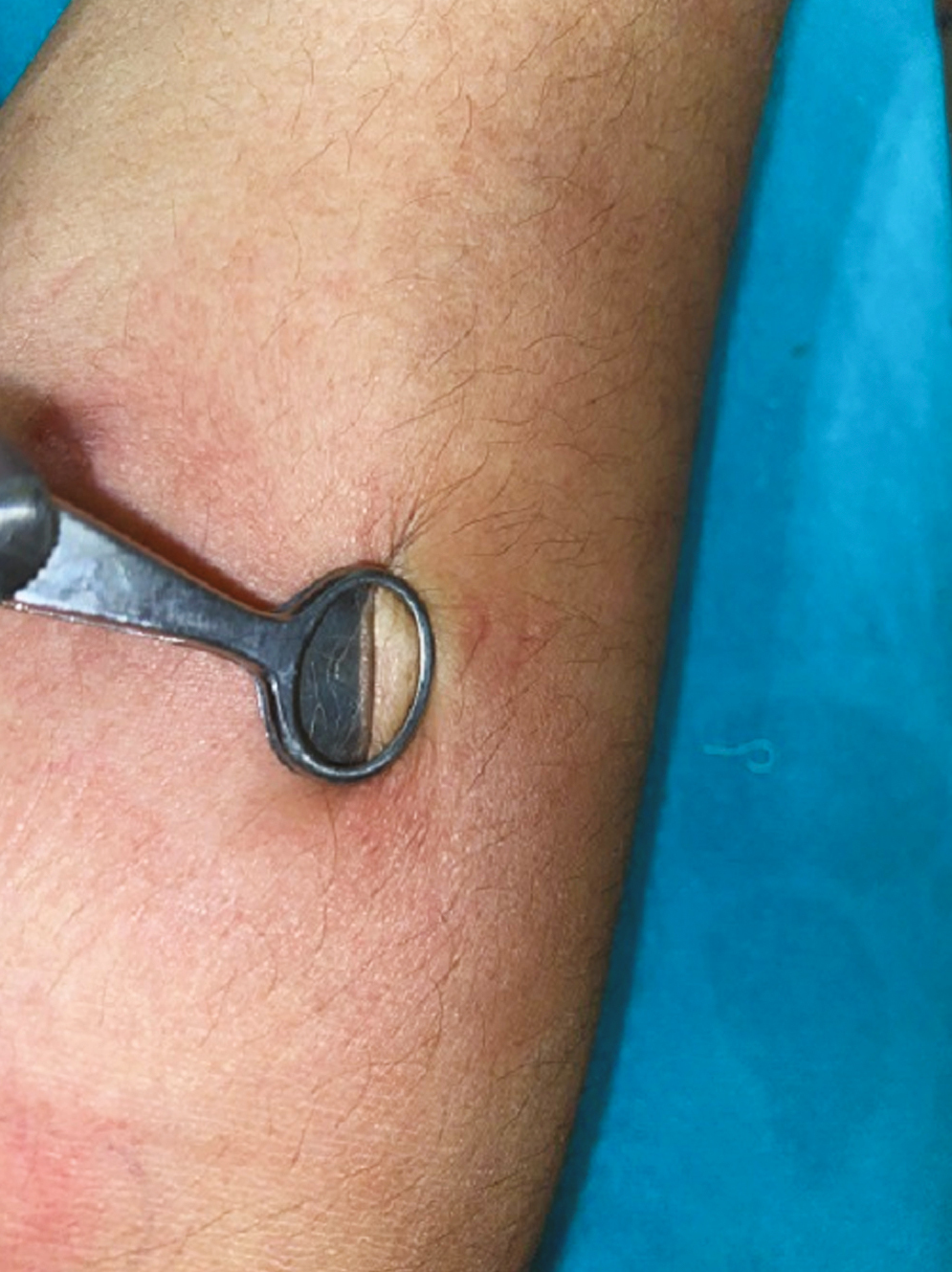
- Slit skin smear over the plaque, creating a avascular zone by chalazion clamp
Financial support and sponsorship
Not applicable.
Conflicts of interest
There are no conflicts of interest.
REFERENCES
- Bloodless nasal alar surgery: Another innovative use of the chalazion clamp. Dermatol Surg. 2009;35:843-4.
- [Google Scholar]
- Use of chalazion clamp for minor salivary gland biopsy in the diagnosis of Sjögren’s syndrome. Surg Cosmet Dermatol. 2009;1:145-6.
- [Google Scholar]
- Textbook on Cutaneous and Aesthetic Surgery. Farmington Hills, MI: JP Medical Ltd; 2012.
- Chalazion clamp in dermatology revisited. Indian J Dermatol Venereol Leprol. 2015;81:280-1.
- [Google Scholar]


