Translate this page into:
Follicular Unit Extraction as a Treatment Modality for Stable Segmental Inguinoscrotal Vitiligo: A Case Report
Address for correspondence: Dr. Yogesh M. Bhingradia, Shivani Skin Care and Cosmetic Clinic, Sarthi Doctor House, Fourth Floor, Hirabah, Varachha Road, Surat, Gujarat, India. E-mail: yogeshbhingradia@gmail.com
This article was originally published by Wolters Kluwer - Medknow and was migrated to Scientific Scholar after the change of Publisher.
Abstract
Abstract
Vitiligo is a common form of autoimmune, localized, or generalized cutaneous depigmentary disorder which has a detrimental effect on psychological and also psychosexual function of many individuals. It is an acquired condition resulting from the progressive loss of melanocytes. Here, we report a case of stable segmental vitiligo affecting inguinoscrotal region, which was successfully treated by follicular unit extraction (FUE).
Keywords
Follicular unit transplantation
inguinoscrotal vitiligo
repigmentation
INTRODUCTION
Vitiligo is an acquired, autoimmune, cutaneous depigmentary disorder affecting any age group. Non-glaberous areas (hair-bearing skin) respond well to the treatment when compared with glaberous area (non-hair-bearing skin) to the medication. Autologous miniature punch grafting and non-cultured melanocyte cellular suspension are well-documented surgery for stable inguinoscrotal vitiligo, but it requires to restrict the mobility of patient for at least 7 days. The acceptance of melanin over the region reduces as growing hair beneath the dressing lifts up the harvested melanocytes, thus reducing its surface contact. Surgical treatment of vitiligo is applicable when medical and phototherapy treatment fails. This case is presented to highlight the safety and efficacy of follicular unit extraction (FUE) in a patient with stable segmental vitiligo over inguinoscrotal region.
CASE HISTORY
A 16-year-old male patient presented with depigmented patch with few perifollicular pigmented macules over the right side of inguinoscrotal region along with single depigmented patch over the medial side of right thigh since 10 years [Figure 1]. The patient was advised multiple topical and oral therapies along with phototherapy treatment for the same with no improvement. The disease remained stable for over 2 years with negative Koebner’s phenomenon. On clinical examination, two depigmented patches of approximately 8 * 5 cm and 3*3 cm were present on the right side of scrotum extending to base of the penis, pubic region, and on the medial aspect of right thigh, respectively, along with positive leukotrichia. A diagnosis of segmental vitiligo was made based on clinical examination. There was no history of any bleeding disorder or keloid formation. Based on the history and clinical examination, we planned a surgical approach using FUE method.
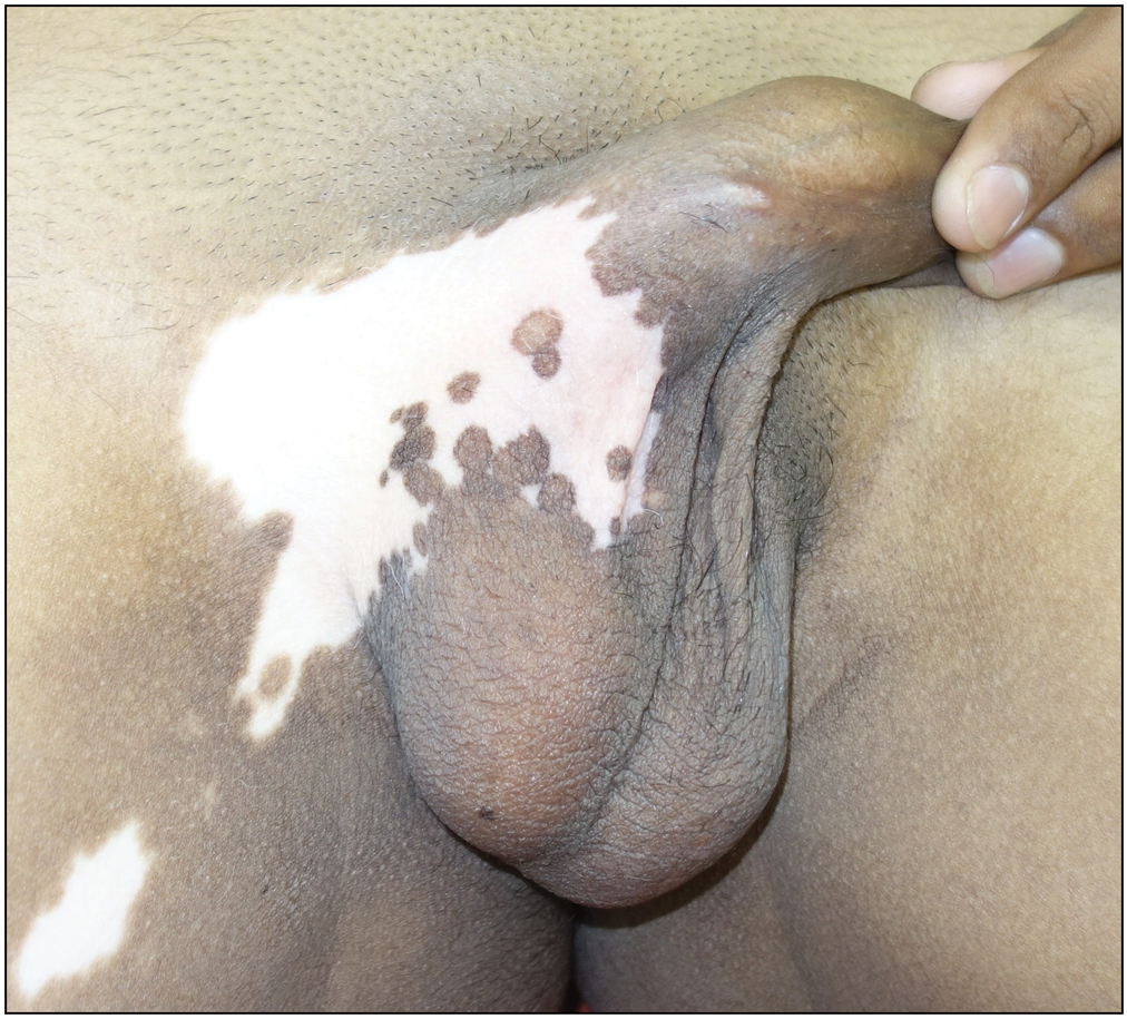
- Pre-procedural picture showing stable vitiligo patch over scrotal and pubic region
PROCEDURE
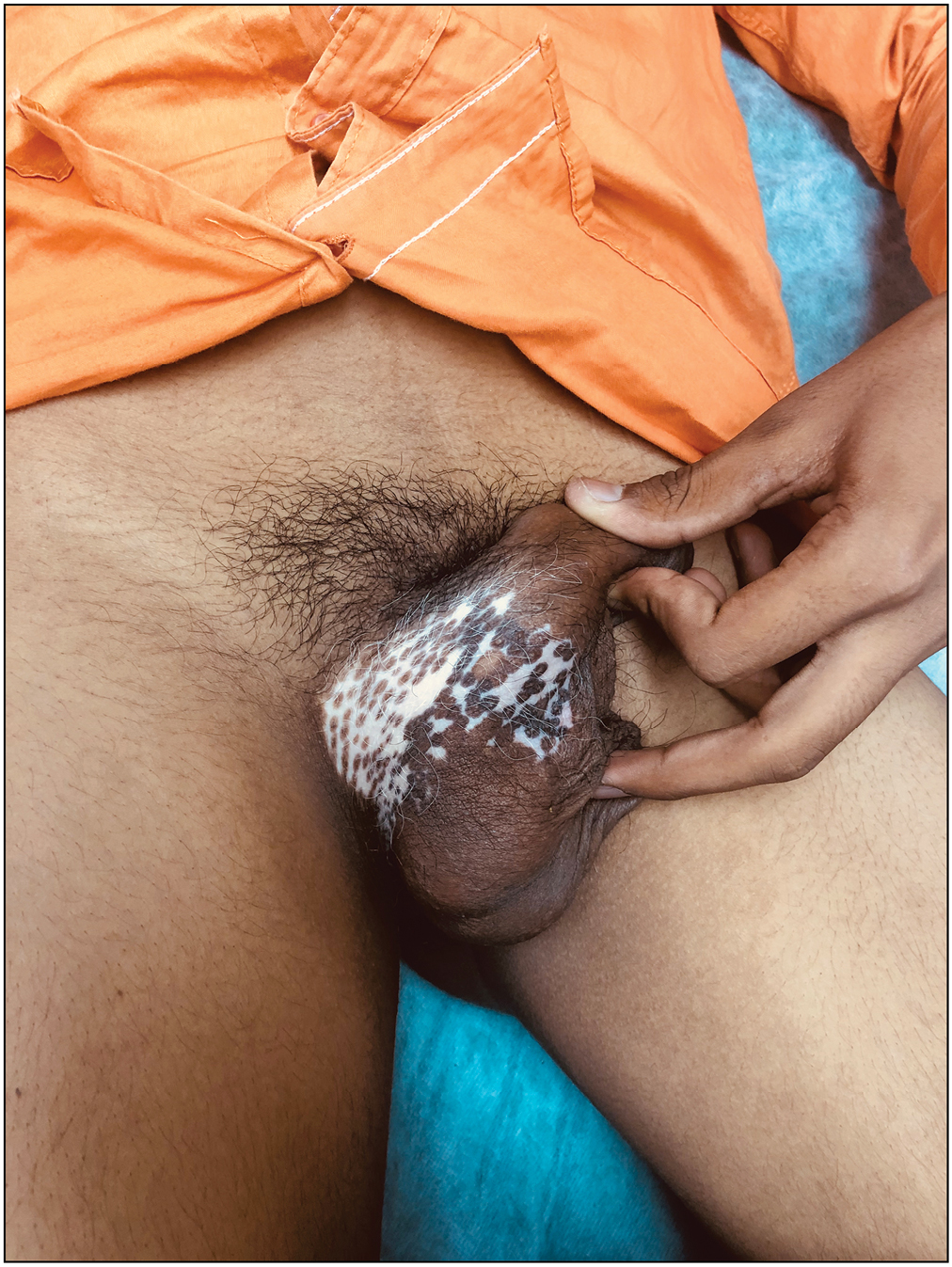
After obtaining an informed, written consent from the patient, the recipient area (occipital area of scalp) and the donor area were shaved, cleaned thoroughly using betadine, spirit, and normal saline [Figure 2]. The graft was harvested using a 0.7 mm micromotor punch after infilterating the area with local anesthesia. The follicular unit grafts were preserved in petri dish containing cold normal saline. After anesthetizing recipient area, slits were made using 18G needle and were then enhanced using gentian violet [Figure 3]. The follicular unit grafts were transplanted into the recipient area using jewel’s forcep. The grafts were fixed using surgical glue to avoid dressing, thus improving the patient’s compliability, immobility, and enhancing the chances of graft acceptance. The patient was started on oral antibiotics for 5 days. Topical tacrolimus and narrow band UVB therapy (NBUVB) were started after 2 weeks of surgery. No immediate or late post-procedure complications were reported. Perifollicular repigmentation was noted at the end of 4 weeks over scrotal skin, whereas depigmented patch over thigh showed complete regimentation in 6 weeks [Figure 4]. Complete pigmentation over scrotal skin was observed in 16 weeks [Figure 5]. Leukotrichia as assumed was not improved with the treatment. No history of any recurrence was noted till date.
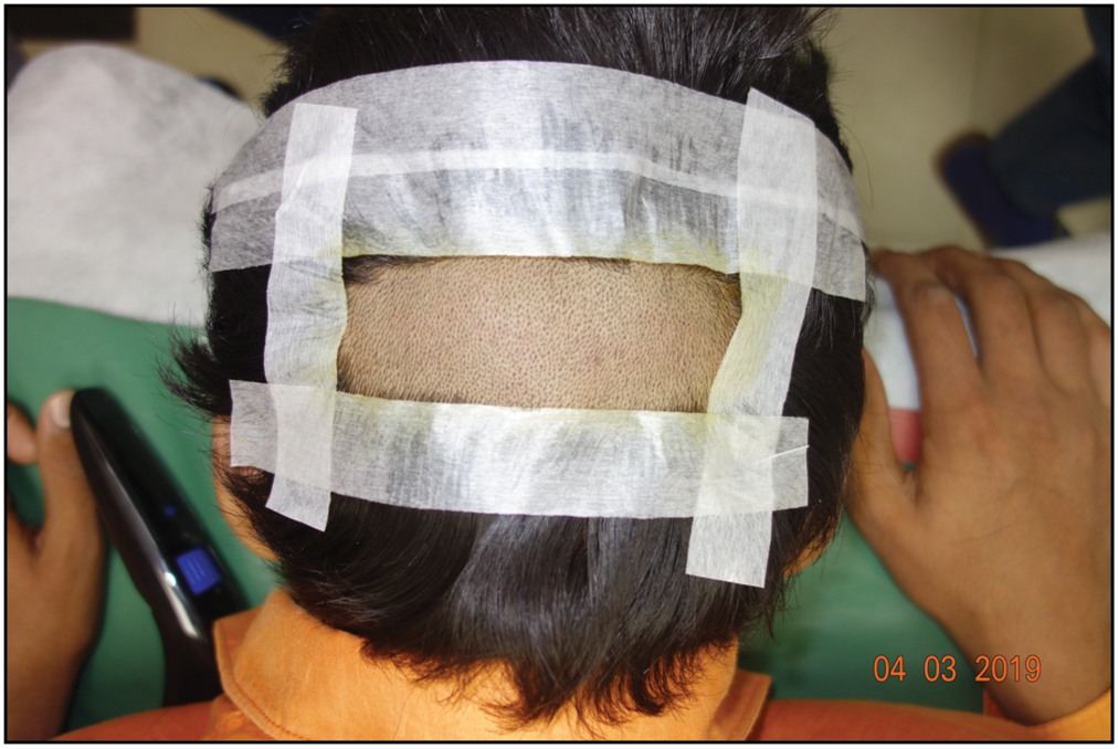
- Follicular units extracted from the occipital region
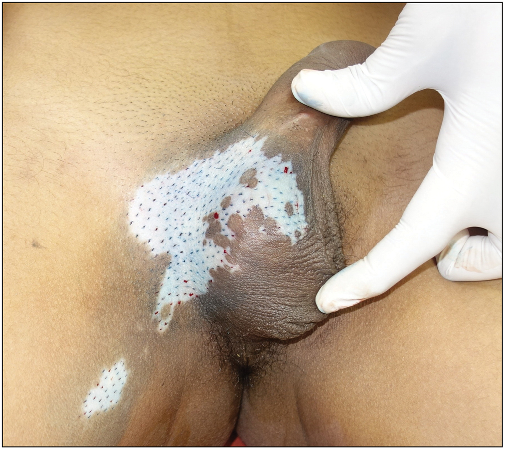
- Visible slits after application of gentian violet over recipient area
- Partial repigmentation after 6 weeks of procedure
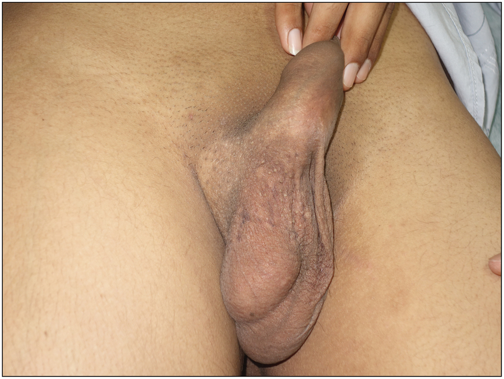
- Complete repigmentation with leukotrichia is seen after 16 weeks of procedure
DISCUSSION
Vitiligo is a process in which only active (melanin-producing) melanocytes are destroyed and the inactive ones in the outer route sheath are preserved and serve as the only source for repigmentation.[1] The genital is a commonly affected site for vitiligo in men, and it is sometimes the only site affected.[2] The treatment of genital vitiligo is quite complex. The existence of undifferentiated stem cells in hair follicle units can serve as a source of melanocytes for repigmentation, which is the rationale behind follicular unit transplantation. As hair follicle melanocytes are more resistant to the vitiligo process,[3] hair restoration in a vitiligo patch may be a good modality of treatment, especially in those with leukotrichia, which are difficult to treat with other methods.
Keeping the above facts in mind, we decided to transplant the hair follicular unit in the patient with stable inguinoscrotal, segmental vitiligo. After follicle transplantation, the peripheral spread of pigmentation ranged from 5 to 12 mm,[4] thus the density of 6–8 follicular/cm2 was sufficient to induce complete repigmentation.
As the occiput is the donor site, we can completely avoid the complications of other grafting procedures (e.g., punch grafting, split-thickness skin grafting, and blister technique) which have visible scar and hypo- or hyperpigmentation at the donor site.[5] Cobble stoning over the recipient site is more with punch grafting than that with follicular transplantation as there are more melanocytes in hair follicle than normal epidermis, and the cause of a more acceptable color match in hair transplantation than in other methods could be related to the stem cell migration from the graft and then location-specific transient that amplifies cells proliferation.[6]
CONCLUSION
Genital segmental vitiligo over inguinoscrotal region remains challenging to treat as pubic region has many preexisting hairs and scrotal skin has sparse fine hairs and also the movement restriction, and dressing over this area is difficult after any surgical procedure. So, we opted for FUE method to avoid the above-mentioned complications.
Declaration of patient consent
The authors certify that they have obtained all appropriate patient consent forms. In the form the patient(s) has/have given his/her/their consent for his/her/their images and other clinical information to be reported in the journal. The patients understand that their names and initials will not be published and due efforts will be made to conceal their identity, but anonymity cannot be guaranteed.
Financial support and sponsorship
Nil.
Conflicts of interest
There are no conflicts of interest.
REFERENCES
- Amelanotic melanocytes in the outer sheath of the human hair follicle. J Invest Dermatol. 1959;33:295-7.
- [Google Scholar]
- Non-veneral penile dermatoses. In: Kumar B, Gupta S, eds. Sexually Transmitted Infections. New Delhi: Elsevier; 2005. p. :539-64.
- [Google Scholar]
- Follicular unit extraction as a therapeutic option for vitiligo. J Cutan Aesthet Surg. 2013;6:229-31.
- [Google Scholar]
- Transplantation of hair follicles for vitiligo. In: Gupta S, Olsson MJ, Kanwar AJ, Ortonne JP, eds. Surgical Management of Vitiligo (1st ed.). Oxford: Blackwell; 2007. p. :123-7.
- [Google Scholar]
- A study of hair follicular transplantation as a treatment option for vitiligo. J Cutan Aesthet Surg. 2015;8:211-7.
- [Google Scholar]
- Follicular unit extraction technique in treating stable vitiligo with leukotrichia. J Dermatology Surg. 2018;22:72-4.
- [Google Scholar]


