Translate this page into:
Retronychia: A Misdiagnosed Cause of Paronychia
Address for correspondence: Dr. Sankey Sana Mariam, Roots Institute of Dermatological Sciences, HRBR 2nd Stage, 5th B Main Kalyannagar, Bengaluru 560043, Karnataka, India. E-mail: sanasankey@gmail.com
This article was originally published by Wolters Kluwer - Medknow and was migrated to Scientific Scholar after the change of Publisher.
Abstract
Abstract
Retronychia refers to the embedding of the nail into the proximal nail fold. Patients present with chronic paronychia in the setting of disrupted nail growth. Nail avulsion is curative and unlike other forms of ingrown nails, it does not tend to recur. We report a case of retronychia who presented with pain and swelling around bilateral great toes. Further examination showed growth of overlapping nail plates, which led to the diagnosis of retronychia. This article emphasizes the clinical features and treatment options available for retronychia, thereby avoiding misdiagnosis.
Keywords
Embedding
nail avulsion
retronychia
retronychia, chronic paronychia

INTRODUCTION
The term “retronychia” was introduced in 1999 by De Berker and Rendall.[1] It is characterized by the embedding of the proximal nail plate into the proximal nail fold and the growth of several nail plates, misaligned beneath each other.[1] This condition is mainly caused by trauma that leads to onycholysis.[2] Due to the similarity with other diseases like onychomycosis and chronic paronychia, it may be challenging to diagnose a case of retronychia. Although it may probably be a common condition, only a few cases have been reported in the literature.
CASE HISTORY
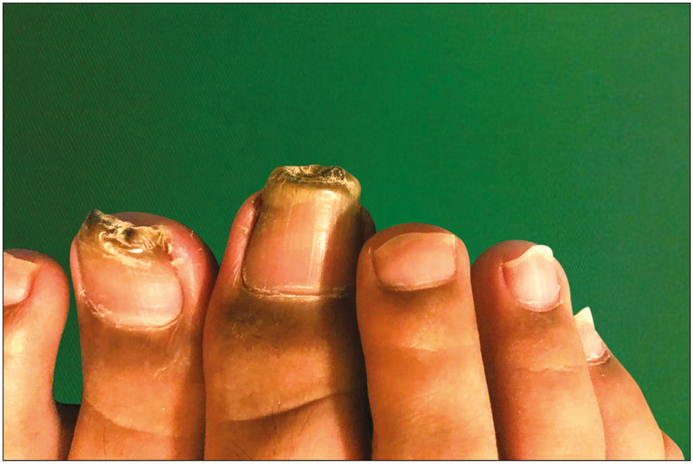
A 15-year-old boy was referred to our OPD with pain and swelling around bilateral great toes, which was present since one and half years. On examination, there was edema, erythema, crusts seen along the periungual skin, proximal nail fold pigmentation and the nail plate showed yellow-to-black discoloration on both halluces. The rest of the nails of both hands and feet were normal [Figure 1]. The patient reported playing for the school football team and wearing shoes on a daily basis. Onychomycosis was excluded based on the negative potassium hydroxide test and fungal culture. Based on the following findings, we concluded the patient presented with retronychia and advised nail avulsion. During nail avulsion, three generations of nail plate in the left hallux [Figure 2] and five generations of nail plate in the right hallux were seen and successfully removed [Figures 3and 4]. The patient had normal growth of nails and was symptom free at 8 months of follow-up [Figure 5].
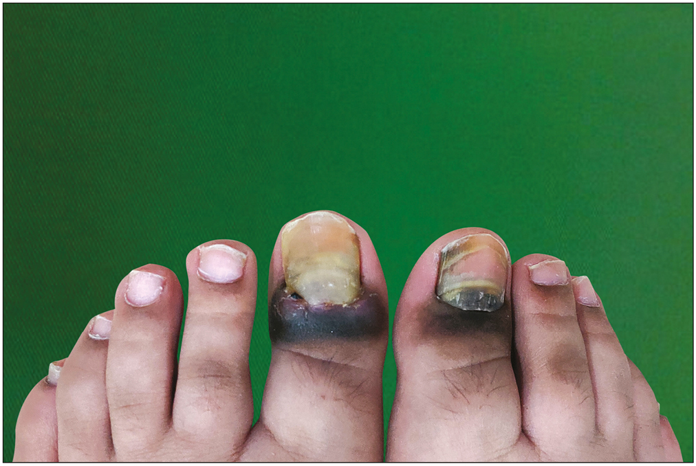
- Chronic paronychia, proximal nail fold pigmentation, and yellow-to-black discoloration of nail plate on both halluces
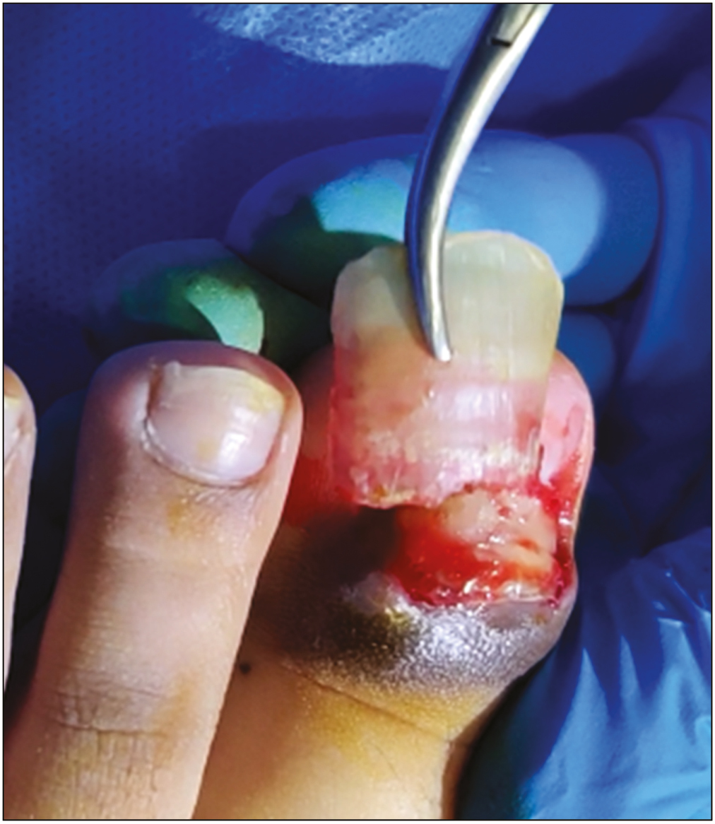
- Nail avulsion of left hallux
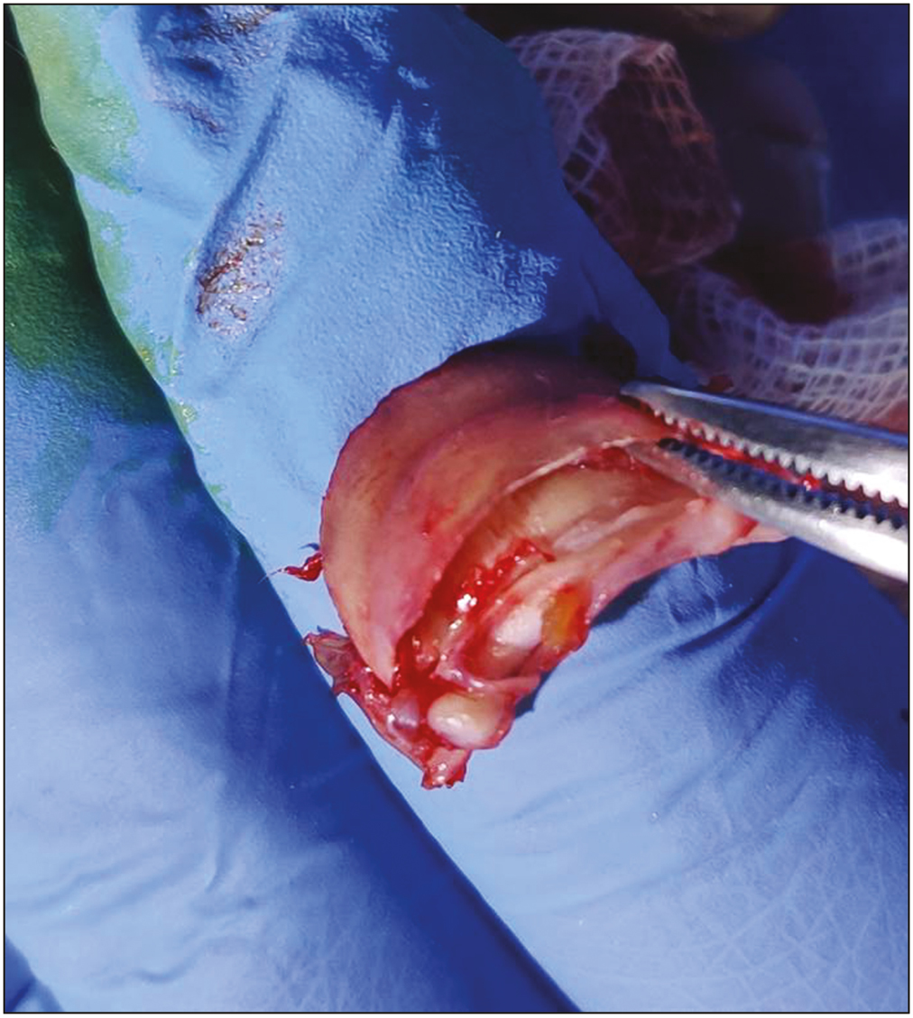
- Right hallux: stacking of three nail plates
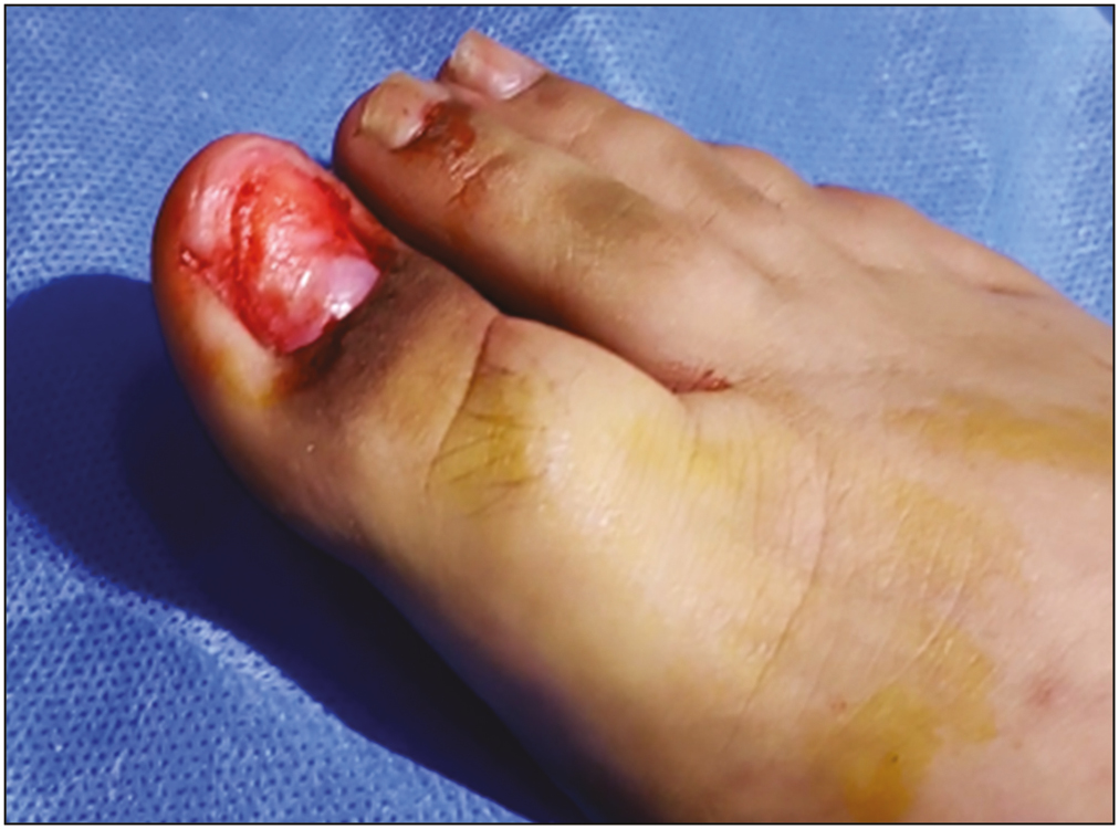
- Right hallux: new nail growth on the nail bed
- Eight-month postoperation
DISCUSSION
Retronychia refers to the embedding of the proximal edge of the nail plate into the proximal nail fold. It is a painful condition that is underdiagnosed due to diverse clinical manifestations.[3] Risk factors include sports activities and the use of heels or ill-fitting shoes. Toenails are more prone to microtrauma and hence are most frequently affected by retronychia.[4] It exhibits a triad of clinical findings that are chronic paronychia, liquid discharge from under the nail fold, and disruption of linear growth of the nail. Other clinical features include granulation tissue, yellowish coloration of the nail plate, and Beau’s lines.[5]
The major pathogenic event in the development of this condition includes several degrees of onycholysis that leads to nail separation from the matrix and the nail bed, which no longer supports the nail’s forward growth. As the nail is no longer attached to the nail bed, it can be pushed back into the nail pocket, injuring the overlying proximal nail fold causing chronic inflammation. In retronychia, the old nail remains in the nail pocket and the new one pushes it up instead of out, increasing the nail thickness proximally.[36] The first stage of retronychia is undiagnosed characterized by an arrest in the nail plate’s growth, accompanied by xanthonychia, presence of exudate under the proximal nail fold and discreet paronychia, and a late stage in which in addition to the aforementioned signs, intense paronychia and an important thickening of the nail plate can be observed.[7]
Three-dimensional ultrasound examination is a noninvasive technique to confirm the diagnosis.[8] It shows a decreased distance between the origin of the nail plate and the base of the distal phalanx, thickening, decreased echogenicity, and increased blood flow in the dermis of the posterior nail fold and proximal nail bed.[9]
Differential diagnoses include candidiasis or bacterial infections, psoriatic arthritis, cysts, and subungual tumors(glomus tumors, cutaneous squamous cell carcinoma, myxoid cyst, Bowen’s disease, keratoacanthoma, and amelanotic malignant melanoma).[610]
Management of retronychia consists of complete avulsion of the nail plate. It is an inexpensive, rapid, and curative procedure following which the pain quickly subsides, and a new nail plate grows back normally. Patients who refuse surgery can be successfully treated with topical steroids or chemical avulsion with 50% urea and 10% salicylic acid in vaseline, white petroleum, covered by occlusive dressing during a week. Consistent taping is a valuable alternative. In the presence of severe pain, surgery is mandatory to free the distal edge of the plate (Dubois or Super “U” techniques). To prevent such a complication, an acrylic nail can be attached to the newly growing nail when it has reached one-third of its length.[611]
Following avulsion recurrences are rare, unlike other forms of incarceration.
CONCLUSION
Retronychia, though common due to its diverse presentation, can be challenging to diagnose. Posttraumatic proximal paronychia, proximal thickening of the nail plate, xanthonychia, and overlapping of several nails as well as negative fungal cultures are key to suspect the condition. Proximal nail avulsion is considered the first-line treatment for retronychia, and recurrences after this approach are extremely rare.
Declaration of patient consent
The authors certify that they have obtained all appropriate patient consent forms. In the form the patient(s) has/have given his/her/their consent for his/her/their images and other clinical information to be reported in the journal. The patients understand that their names and initials will not be published and due efforts will be made to conceal their identity, but anonymity cannot be guaranteed.
Financial support and sponsorship
Nil.
Conflicts of interest
There are no conflicts of interest.
REFERENCES
- Retronychia-clinical and pathophysiological aspects. J Eur Acad Dermatol Venereol. 2016;30:16-19.
- [Google Scholar]
- Risk factors, clinical variants and therapeutic outcome of retronychia: A retrospective study of 18 patients. Eur J Dermatol. 2016;26:377-81.
- [Google Scholar]
- Retronychia: A common and misdiagnosed condition. Asian J Res Dermatol Sci. 2021;4:19-24.
- [Google Scholar]
- Ultrasonographic criteria for diagnosing unilateral and bilateral retronychia. J Ultrasound Med. 2018;37:1201-9.
- [Google Scholar]
- Finger retronychias detected early by 3d ultrasound examination. J Eur Acad Dermatol Venereol. 2012;26:254-6.
- [Google Scholar]
- Surgery of the nail plate. In: Richert B, Di Chiacchio N, Haneke E, eds. Nail Surgery. London: Healthcare; 2010. p. :32.
- [Google Scholar]


