
Translate this page into:
A supraciliary subcutaneous browlift for tattooed eyebrow ptosis

*Corresponding author: Gabriela Esther Beraja, Department of Plastic Surgery, Beraja Medical Institute, Florida, United States. gaby@beraja.co
-
Received: ,
Accepted: ,
How to cite this article: Beraja GE, Beraja V. A supraciliary subcutaneous browlift for tattooed eyebrow ptosis. J Cutan Aesthet Surg. doi: 10.25259/jcas_167_23
Abstract
Patients with eyebrow ptosis often remove their natural eyebrow hairs and tattoo the skin above the brow in their desired position. Frequently, these tattooed brows are drawn significantly higher than their actual anatomical location, resulting in an artificial appearance. By extending the incision laterally into the temporal region and meticulously undermining the subcutaneous tissue beneath the eyebrow, supraciliary, and temporal regions, we can effectively correct ptosis and achieve the creation of more esthetically pleasing tattooed brows.
Keywords
Supraciliary subcutaneous brow lift
Tattooed eyebrows
Eyebrow ptosis

INTRODUCTION
In an ideal eyebrow, the innermost point of the brow aligns with the outer edge of the nose (ala) and the inner corner of the eye (inner canthus). The outermost part of the brow should extend along an angled line originating from the farthest point of the ala through the outer corner of the eye (lateral canthus). In addition, the inner and outer ends of the brow should roughly align at the same height. It has been reported that the arch of the brow in a woman should be positioned vertically above the outer edge of the lateral limbus.1
As we age, the natural connections between the eyebrow and the periosteum weaken. This weakening causes the eyebrow to shift downward, requiring the recruitment of the frontalis muscle to lift the eyebrow. The lateral aspect of the eyebrow lacks deep structural attachments from the periosteum and fibers from the frontalis muscle do not extend to the lateral brow, meaning that even with maximal contraction of the frontalis muscle, sagging of the lateral eyebrow persists and may even encroach on the space of the eyelid forming a lateral hood.2
Patients with drooping eyebrows often epilate the eyebrow hairs and tattoo the skin above the eyebrow in the location that they desire the eyebrow to be. Many patients position their brows much higher than their actual anatomical location, creating an unnatural appearance. Our proposed technique closely aligns with the direct brow lift approach outlined by Karimi et al.3 However, we modified it by extending the incision laterally into the temporal region and conducting precise undermining of the subcutaneous tissue beneath the eyebrow, supraciliary, and temporal regions.
SURGICAL TECHNIQUE
While the patient is sitting upright, draw a line marking the superior row of the eyebrow follicles from medial to lateral with a surgical skin marking pen. Extend the lateral marking 1–1.5 cm toward the temporal area at an oblique angle following the relaxed skin tension lines by gently pinching the skin and identifying the skin creases. Digitally elevate the lateral portion of the eyebrow to the desired level, and place the marking pen near, but not touching the superior brow border. Once the brow is released, mark the skin beneath the pen tip to represent point “A,” usually 1–1.5 cm above the eyebrow. Mark the skin forming a gentle curve from the medial to the lateral end of the initial line passing through point “A” [Figure 1a]. The skin is prepped and draped sterile. Infiltrate the subcutaneous tissue with 2% lidocaine and 1:100,000 epinephrine solution extending the infiltration 0.5 cm superiorly and 1–1.5 cm inferiorly beyond the marking to anesthetize the area to be undermined and elevated. Remove the skin in the subcutaneous plane and cauterize capillaries with a bovie. Dissect in the subcutaneous plane the area that was anesthetized and cauterized capillaries with the bovie [Figure 1b]. The dissection is superficial to the orbicularis muscle. The skin is closed in 2 layers; a deep layer of buried running absorbable suture for the subcutaneous tissue and a 6–0 nylon for the dermis [Figure 1c]. By applying the techniques outlined above, tattooed eyebrows that appear unnaturally positioned [Figure 2] can be corrected to enhance their aesthetic appeal [Figure 3].
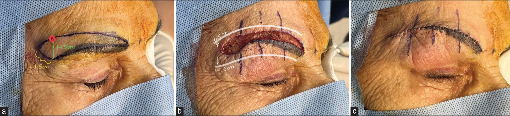
- Surgical steps. (a) Creating point “A” and drawing your lines, (b)Undermining technique above and below, (c) Closing the skin.
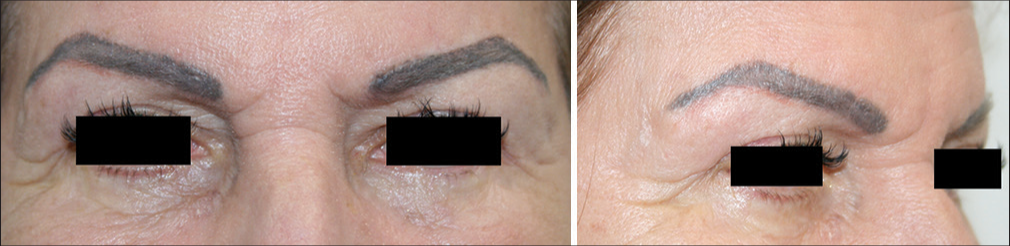
- Before surgery.
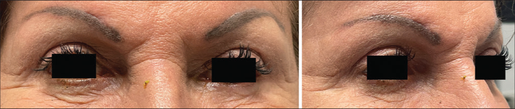
- After surgery (5 months).
Learning Points
Patients with eyebrow ptosis often remove their natural eyebrow hairs and tattoo the skin above their brows in the desired position
The eyebrow tattoo is often placed significantly higher than its true anatomical position, resulting in an unnatural look
By extending the incision laterally into the temporal region and meticulously undermining the subcutaneous tissue beneath the eyebrow, supraciliary, and temporal regions, we can effectively correct ptosis and create more esthetically pleasing tattooed brows
This approach positions the tattooed brows in a more natural location and corrects eyebrow ptosis.
The lateral hooding also shows significant improvement.
CONCLUSION
Many patients with drooping eyebrows often epilate the eyebrow hairs and tattoo the skin above the eyebrow in a position that is much higher than the proper anatomical location, creating an unnatural appearance. By extending the incision laterally into the temporal region and conducting precise undermining of the subcutaneous tissue beneath the eyebrow, supraciliary, and temporal regions, it is possible to correct the ptosis and create more esthetically pleasing tattooed brows. The lateral hooding showed a remarkable improvement; thus, this method can be used in droopy eyebrows without tattoos as well. Furthermore, this technique offers the possibility of deeper fixation of the newly created eyebrow position to the periosteum. We have applied this technique to patients with skin of color, and their satisfaction levels have been comparable to those with lighter skin types, yielding equivalent results.
Authors’ contributions
Both the authors, Gabriela E. Beraja and Victor Beraja contributed to the writing and editing of the manuscript.
Ethical approval
Institutional Review Board Approval is not required.
Declaration of patient consent
The authors certify that they have obtained all appropriate patient consent.
Conflicts of interest
There are no conflicts of interest.
Use of artificial intelligence (AI)-assisted technology for manuscript preparation
The authors confirm that there was no use of artificial intelligence (AI)-assisted technology for assisting in the writing or editing of the manuscript and no images were manipulated using AI.
Financial support and sponsorship
Nil.
References
- Aesthetic analysis of the ideal eyebrow shape and position. Eur Arch Otorhinolaryngol. 2016;273:305-10.
- [CrossRef] [Google Scholar]
- Techniques of eyebrow lifting: A narrative review. J Ophthalmic Vis Res. 2020;15:218-35.
- [CrossRef] [Google Scholar]


