
Translate this page into:
Ionized plasma jet therapy for treatment of vascular tumor over glans penis

*Corresponding author: Ajay Dodeja, Department of Dermatology, Narendra Kumar Prasadrao Salve Institute of Medical Sciences and Research Centre and Lata Mangeshkar Hospital, Hingna, Nagpur, India. ajaydodeja1995@gmail.com
-
Received: ,
Accepted: ,
How to cite this article: Dodeja A, Pande S, Prakashey AR. Ionized plasma jet therapy for the treatment of vascular tumor over glans penis. J Cutan Aesthet Surg. doi: 10.25259/JCAS_103_2024
Dear Editor,
There is a broad spectrum of vascular proliferations occurring on genitalia, which are prone to trauma and irritation. We encountered an erythematous, friable nodule that bled on touch, which was thought to be either a genital wart or a vascular tumor. Such slow-growing lesions can cause discomfort, occasional bleeding, and pain, thereby reducing the quality of life. Standard treatments include surgical excision, cryotherapy, or laser therapy; however, these approaches can lead to complications such as recurrence, scarring, and post-procedure discomfort. Ionized plasma jet therapy (IPJT) is an emerging, minimally invasive modality that provides favorable cosmetic outcomes with minimal risk of scarring. In this report, we present a case where IPJT was successfully implemented to treat vascular growth on the glans penis, with cosmetically optimal results and no recurrence at follow-up.
A 42-year-old male presented with a 7 mm sized exophytic, filiform, friable nodule on the glans penis. [Figure 1a] The lesion, which had progressively enlarged over 2 weeks, was prone to bleeding with trivial trauma. He denied any history suggestive of previous trauma, sexually transmitted infections, promiscuity, significant underlying systemic conditions, or any other high-risk behavior. On examination, the lesion was soft, erythematous, and bled on touch. Mucoscopy demonstrated vasculature of undifferentiated morphology in diffuse arrangement pattern within the dark red lacunae, reddish-white homogenous areas on top of the projections suggestive of the vascular tumor, probably thrombosed mucosal wart or pyogenic granuloma.1 There were no features suggestive of any malignant changes.
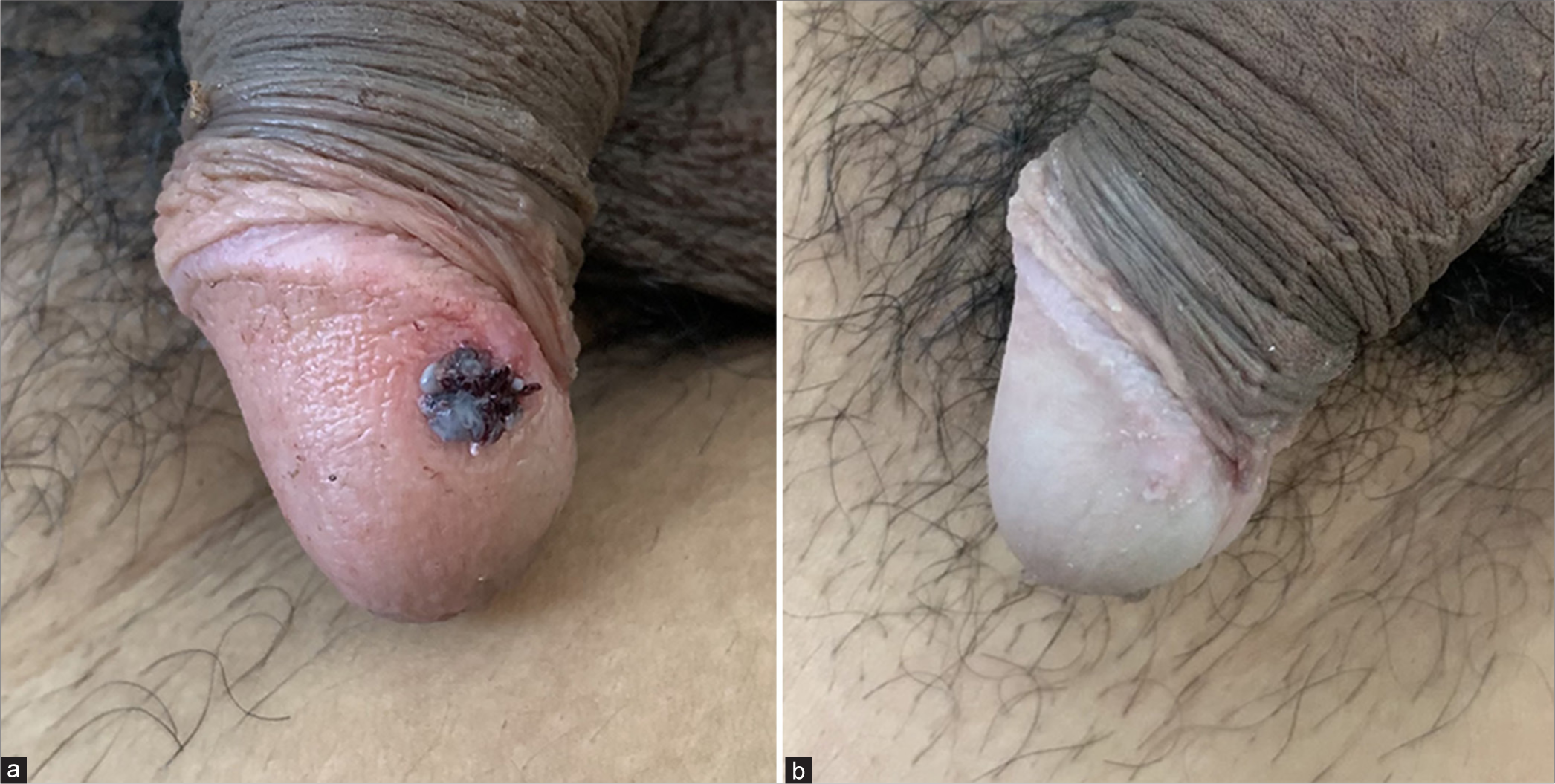
- (a) 7 mm sized exophytic, filiform, and friable nodule on the glans penis (pre-procedure). (b) Healed lesion site without scarring or pigmentary changes (2 weeks post-procedure).
Given the lesion’s location, the patient was apprehensive about the potential for scarring and functional impairment with traditional treatment options. After discussing the risks and benefits of various modalities, the patient opted for IPJT due to its non-invasive nature, reduced risk of hemorrhage and favorable cosmetic outcomes.
The procedure was performed under topical anesthesia. IPJT device was set to a low energy level to avoid damage to the delicate mucosa of the glans penis. The lesion was ablated in a “top to bottom” fashion, targeting one lobule at a time until a thin, flat, and grayish desiccated layer was formed. This ensured immediate coagulation of the lesion. The patient experienced minimal discomfort, and there was minimal intraoperative bleeding. By the end of the procedure, the lesion had visibly ablated, and no further intervention was needed.
At the first follow-up 1-week post-procedure, the treated area showed good healing. The lesion had completely regressed with no signs of infection, bleeding, or significant inflammation. Most notably, the cosmetic outcome was considered optimal, as the glans penis showed no visible scarring or discoloration [Figure 1b].
By the 6th-week follow-up, the area remained lesion-free with complete resolution. The patient was highly satisfied with the treatment results, reporting no functional impairment. A final follow-up 3 months after the procedure confirmed no recurrence of the lesion, further demonstrating the effectiveness of IPJT in this case.
There is a broad spectrum of benign vascular lesions which can occur over the glans penis, and almost all of them cause similar discomfort to the patient. This has a major impact on the self-confidence of the patient. Our provisional diagnosis was genital wart, but pyogenic granuloma could not be ruled out.2 After a brief discussion with the patient, due to his cosmetic and functional concerns, a decision to use IPJT was made since both lesions can be therapeutically cauterized. Surgical excision is routinely chosen as it not only reduces the risk of recurrence but also provides a sample for histopathological examination. However, there is a potential risk of complications such as scarring, pigmentation, dehiscence, and secondary infection. In concordance with modalities such as cryotherapy, electrofulguration and diathermy coagulation, it is not possible to obtain a tissue sample for histopathology with IPJT as well. None of the cases report the use of IPJT for the treatment of genital lesions. In our case, IPJT provided a cosmetically pleasing outcome without compromising treatment efficacy.
The mechanism of IPJT can be explained by the application of direct current in the “space” between the patient’s skin and the equipment’s emitting tip. Due to this, the surrounding atmospheric air is exposed to high voltages, which create conductive electric currents3 and the formation of reactive oxygen species (ROS), reactive nitrogen species, and other gases (OH, NO, and O3).4,5 These gases have antibacterial and protective effects on tissues, leading to the production of controlled thermal lesions on the skin’s superficial layers. Interestingly, the humidity in the air can enhance the plasma jet’s ability to produce ROS.6
IPJT provides a suitable, minimally invasive treatment option for penile vascular tumors. To our knowledge, this is the first case to demonstrate the use of IPJT for the treatment of genital vascular lesions. In our case, IPJT resulted in a cosmetically optimal outcome without visible scarring and no recurrence at the end of 3 months. With this case, we attempt to highlight the potential of IPJT as an alternative to traditional treatments for vascular lesions in cosmetically and functionally critical regions.
Authors’ contributions
Dr Ajay Dodeja, Dr. Sushil Pande and Dr. Arjun Prakashey had full access to all of the data in the study and take responsibility for the integrity and accuracy of the data; study concept and design: Arjun Prakashey; Acquisition, analysis, and interpretation of data: Ajay Dodeja; Drafting of the manuscript: Ajay Dodeja; Critical revision of the manuscript for important intellectual content: Sushil Pande.
Ethical approval
Institutional Review Board approval is not required.
Declaration of patient consent
The authors certify that they have obtained all appropriate patient consent.
Conflicts of interest
There are no conflicts of interest.
Use of artificial intelligence (AI)-assisted technology for manuscript preparation
The authors confirm that there was no use of artificial intelligence (AI)-assisted technology for assisting in the writing or editing of the manuscript and no images were manipulated using AI.
Financial support and sponsorship: Nil.
References
- Pyogenic granuloma of the penis: An uncommon lesion with unusual presentation. Curr Urol. 2017;9:216-8.
- [CrossRef] [PubMed] [Google Scholar]
- Plasma medicine: Applications of cold atmospheric pressure plasma in dermatology. Oxid Med Cell Longev. 2019;2019:3873928.
- [CrossRef] [PubMed] [Google Scholar]
- Characterization of plasma jet equipment used in the treatment of aesthetic affections. Int J Adv Res. 2018;6:595-604.
- [CrossRef] [Google Scholar]
- Identification of the biologically active liquid chemistry induced by a nonthermal atmospheric pressure plasma jet. Biointerphases. 2015;10:29518.
- [CrossRef] [PubMed] [Google Scholar]
- Plasma jet versus electrocarbonization in the treatment of wrinkles of the upper palpebral region. J Clin Aesthet Dermatol. 2024;17:33-40.
- [Google Scholar]


