
Translate this page into:
Balanitis of Zoon treated with 30% salicylic acid lotion – A case series
*Corresponding author: Vikrant Saoji, 22, Dandige Layout, Shankar Nagar, Nagpur, Maharashtra, India. vikrantsaoji@hotmail.com
-
Received: ,
Accepted: ,
How to cite this article: Saoji V, Kulkarni S. Balanitis of Zoon treated with 30% salicylic acid lotion – A case series. J Cutan Aesthet Surg. doi: 10.25259/jcas_171_23
Abstract
Balanitis of Zoon or balanitis circumscripta plasmacellularis is a non-venereal balanitis affecting middle-aged uncircumcised men. It follows a benign protracted course with a minimal tendency for spontaneous resolution. Chronicity of disease and genital distribution is sufficient to evoke anxiety among subjects and is the primary reason for dermatological consultation. Many topical modalities (antibiotics, antifungals, and immunomodulators) have been resorted to as therapeutic measures with variable and unsatisfactory results and recurrences. Circumcision is a definitive and curative treatment. However, 30% salicylic acid (SA) lotion can prove to be a promising therapeutic alternative, especially for subjects reluctant for circumcision. We are presenting a case series of 10 patients with balanitis of the Zoon treated with 30% SA lotion.
Keywords
Balanitis
Salicylic acid
Pseudo frosting

INTRODUCTION
Zoon’s balanitis or plasma cell balanitis is a non-venereal inflammatory dermatosis affecting the glans and prepuce mostly seen in middle-aged uncircumcised men.1
It presents as a persistent shiny red area on the genital area. It follows a chronic, slowly progressive, and mostly asymptomatic course, commonly misdiagnosed and erroneously treated as a sexually transmitted disease or premalignant condition due to genital involvement. The persistent nature of the lesions associated with a condition with a minimal tendency of spontaneous resolution is sufficient to evoke anxiety among patients seeking dermatology consultation. Many topical therapeutic agents are commonly employed for treatment, including topical corticosteroids, calcineurin inhibitors like tacrolimus, and topical immunomodulators like imiquimod, but the response to the treatment remains unsatisfactory. Circumcision has been described as a curative procedure.
We are presenting a case series of 10 patients with Zoon’s balanitis treated with the application of salicylic acid (SA) 30% lotion for its therapeutic peeling effect.
CASE SERIES
10 patients (uncircumcised males) within the age range of 45–80 years, who were clinically diagnosed with Zoon’s balanitis, reluctant to undergo therapeutic circumcision and not responding to conventional topical and systemic treatment modalities (topical and oral antifungal agents and topical steroids) visiting a private dermatology clinic in Nagpur (Central India) for 2 years were included for this case series. A brief overview of the patient profile, clinical presentation, treatment, and follow-up assessment has been depicted in Table 1.
| Patient profile age/duration of lesion | Treatment | Observations and follow-up |
|---|---|---|
| 50-years-male/Lesion since 2 years | 30% SA application 3 sessions, 3 months apart | After 2 sessions, erythema reduced with partial resolution 3rd follow-up resolved with no recurrence after 6 months of follow-up |
| 54-years-male/Lesion since 5 years | 3 sessions of 30% SA application 3 months apart | Lesions cleared after 3rd session no recurrence after 6-month follow-up |
| 72-years-male/Lesion since 5 years | 3 Sessions of 30% SA application 3 months apart | Partial resolution after 3 sessions and reappearance of the lesion after 6 months follow-up |
| 55-years-male/Lesion since 1 year | 30% SA application single session | Lesion cleared in a single session with no recurrence after 6-month follow-up |
| 64-years-male/Lesion since 1 year | 30% SA application single session | Lesion cleared in a single session with no recurrence after 6-month follow-up |
| 79-years-male/Lesion since 2 years | 3 Sessions of 30% SA application 3 months apart | Local ulceration at the site of application developed after 3rd session healed with topical antibiotic treatment no recurrence after a 6-month follow-up |
| 61-years-male/Lesion since 1 year | 3 sessions of 30% SA application 3 months apart | Lesions cleared after 3rd session no recurrence after 6-month follow-up |
| 48-years-male/Lesion since 1 year | 3 sessions of 30% SA application 3 months apart | Lesions cleared after 3rd session no recurrence after 6-month follow-up |
| 74-years-male/Lesion since 3 years | 2 sessions of 30% SA application 3 months apart | Partial resolution after 1stsession complete resolution after 2nd session recurrence observed after 6 months |
| 57-years-male/Lesion since 2 years | A single session of 30% SA application | Lesions cleared in a single session no recurrence was observed after 6-month follow-up |
SA: Salicylic acid
Out of the total 12 patients recruited initially, 2 cases aged 55 and 65 years each were excluded as they did not turn up for subsequent follow-up after the first session of the SA application. Thus, 10 cases were continued for follow-up and results.
Those with multiple sexual partners and a history of treatment for other sexually transmitted diseases were excluded.
All patients presented with a moist, shiny, asymptomatic, erythematous patch on the glans or corona glandis without any inguinal lymphadenopathy, with an average duration range spanning 1–3 years
All of them had received oral and topical antifungals and steroids for variable durations with minimal or no improvement.
Based on their clinical profile, examination, and poor response to conventional treatment regimens targeting differential diagnoses of balanitis, they were diagnosed clinically as refractory variants of Zoon’s balanitis.
After taking adequate informed consent, 30% SA lotion (a freshly prepared SA solution in acetone) was applied about 0.5–1 mL on the affected area. The urethral orifice was safeguarded by a topical Vaseline application. Pseudofrosting was witnessed only in the inflamed area after the SA application.
Cold compresses were applied immediately after SA application to the affected area to minimize the burning sensation. SA was used as monotherapy, and no other treatment modality was employed as a combination regimen in these patients. Patients were evaluated every 3 months with partial or non-resolution of lesions; a similar procedure was repeated every 3 months for a maximum of 3 sessions. With complete resolution of lesions during any session or partial resolution even after 3 sessions, further treatment was discontinued. Patients were asked to maintain good hygiene and were allowed to apply topical moisturizers only for 6 months and follow up for the same duration for any signs of recurrence or complications of SA application.
Observations
In 5 patients, the lesion cleared after 3 sessions of 30% SA application every 3 months [Figures 1-6]. 1 patient (79 years male) of these 5 developed local ulceration which healed with topical antibiotic application with clearance of the lesion. 1 patient aged 72 years reported partial resolution with residual erythema at the end of 3 sessions and the reappearance of moist plaque after a 6-month surveillance period. Another patient aged 74 years initially reported complete resolution within 2 sessions but subsequently reported recurrence after 6-month follow-up.
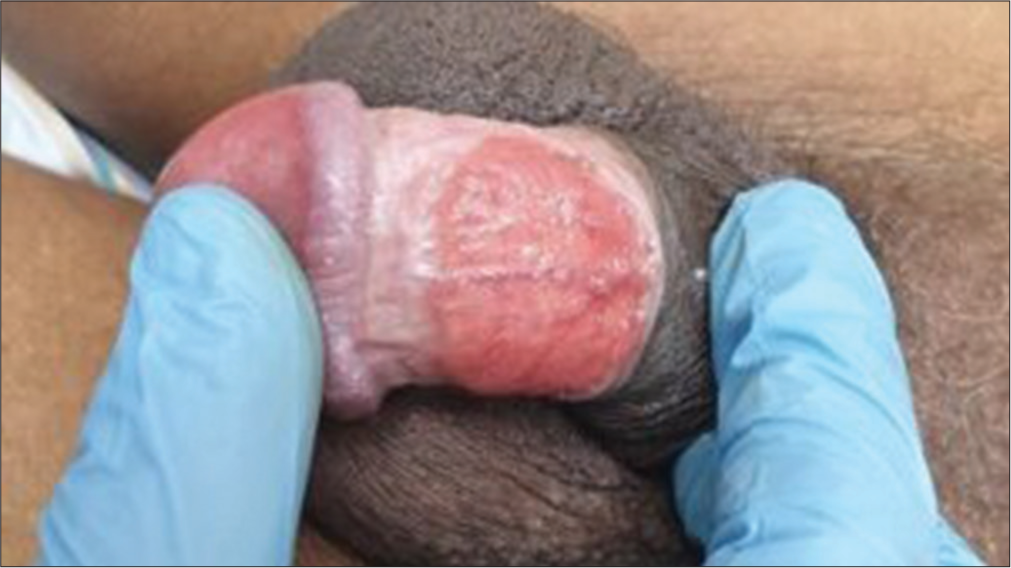
- 50-year-male with lesions for 2 years; Pre-treatment.
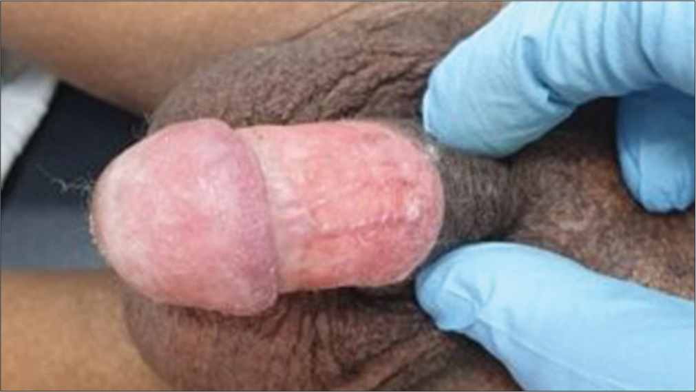
- Result after 3 sessions of 30% salicylic acid application.
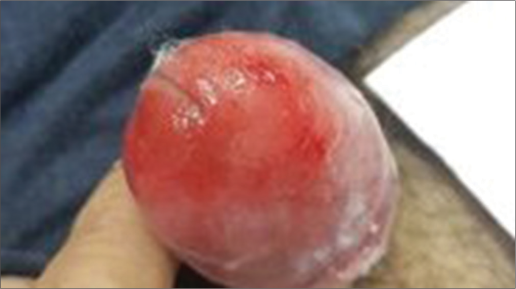
- 48-year-old male with lesion for 1 year; Pre-treatment.
- Partial improvement after 2 sessions of 30% salicylic acid application.
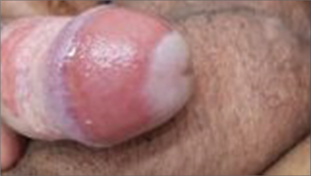
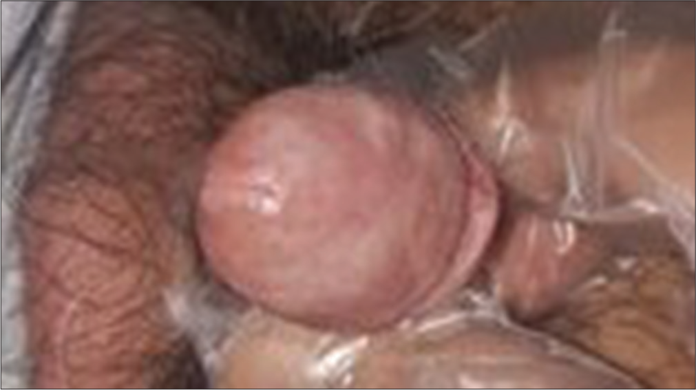
- 55-year-old male with lesion for 2 years; Pre-treatment.
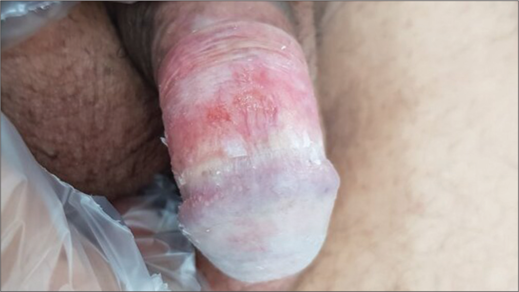
- Result after single session of 30 % salicylic acid application.
3 patients (age 55 years, 57 years, and 64 years) reported resolution with a single session of 30% SA application with no recurrence during surveillance
DISCUSSION
Zoon’s balanitis was highlighted as a distinct subset of the list of differential diagnoses of non-venereal balanitis by Zoon in 1952.1,2
Although the entity is benign with insidious onset and a slowly protracted chronic non-remitting course, less awareness about the entity and its distribution involving genitals is alarming for most patients to seek dermatological consultation.
The etiology is not conclusively established, but poor hygiene with smegma accumulation, chronic local infection, friction, and a tropical climate with excessive moisture are hypothesized to be some of the well-known triggers. Complete resolutions or long-term remissions have been observed post-circumcision.3
Clinically, it most commonly presents as a circumscribed, shiny, moist red plaque affecting the glans penis, though the sub preputial area and coronal sulcus may also be involved. Usually asymptomatic, a few patients may report mild pruritus or burning sensations.1
Histologically, during different phases of evolution, it reveals epidermal changes like acanthosis (with diamond-shaped or lozenge keratinocytes in the lower spinous layer), parakeratosis, and spongiosis, along with characteristic dermal features like a pleomorphic band-like inflammatory infiltrate at the dermo-epidermal interface with a predominance of plasma cells (more than 50%), along with neutrophils, macrophages, lymphocytes, and eosinophils.2,3
Zoon’s balanitis needs to be distinguished from an array of differential diagnoses that closely simulate this entity. Common among them include candidal balanoposthitis, erythroplasia of queyrat, genital psoriasis, lichen planus, Reiter’s disease, allergic/irritant contact dermatitis, fixed drug eruptions, etc.
The most common therapeutic modalities for the treatment of Zoon’s balanitis include mild-to-midpoint topical corticosteroids, topical calcineurin inhibitors (tacrolimus and Pimecrolimus), topical antibiotics (fusidic acid, mupirocin), and topical immunomodulators like imiquimod.4-8 The preferred surgical modality recommended is circumcision.9 Previous studies document the use of non-surgical energy-based ablative modalities like CO2 lasers with variable results.10
Although all these therapeutic options have been used as monotherapy or in various permutations, they have not fared well in achieving clinical remission or complete resolution of the condition.
The etiology is unknown, but most probably it is a chronic, reactive, principally irritant mucositis. Thereby, physical destruction of the overlying epithelium may be effective, thus envisaging the need for exploring chemical ablative modalities like SA for the treatment.
Topical SA, when applied, is actively taken up by inflamed tissue as is observed by prominent pseudofrosting in lesional skin compared to perilesional skin. 30% of SA has a peeling or exfoliative action with subsequent regenerative healing and clearance of the lesion.
Probable speculative mechanisms of action of SA in inducing remission or resolution of lesions of Zoon’s balanitis include sloughing off the denuded or inflamed area of the epidermis by inducing full-thickness acute epidermal injury and peeling action: mobilization and reorganization of the existing chronic inflammatory framework (plasma cells and macrophages) to the acute inflammatory pool (neutrophils), which accelerates the process of wound healing.11-14
CONCLUSION
Although initiated as a small-scale pilot project, the results of the topical SA 30% formulation in the treatment of Zoon’s balanitis appeared promising and provided new insights into widening therapeutic horizons, especially inpatient groups reluctant to circumcision. Nonetheless, large-scale studies are warranted with the recruitment of a substantial number of patients and prospective surveillance for a longer duration before extrapolating its therapeutic efficacy to the general population and duly incorporating it as a part of standard treatment protocols.
Authors’ contributions
Sandeep Kulkarni and Vikrant Saoji participated in Patient selection, treatment protocol formulation, actual treatment, follow up evaluation,data compilation and manuscript preparation.
Ethical approval
Institutional Review Board approval is not required as the study was a pilot study conducted in a private clinic with a very small sample size.
Declaration of patient consent
The authors certify that they have obtained all appropriate patient consent.
Conflicts of interest
There are no conflicts of interest.
Use of artificial intelligence (AI)-assisted technology for manuscript preparation
The authors confirm that there was no use of artificial intelligence (AI)-assisted technology for assisting in the writing or editing of the manuscript and no images were manipulated using AI.
Financial support and sponsorship
Nil.
References
- Chronic benign circumscript plasmocytic balanoposthitis. Dermatologica. 1952;105:1-7.
- [CrossRef] [Google Scholar]
- Dermatoses of the male genitalia In: Griffiths C, Barker J, Bleiker T, Chalmers R, Creamer D, eds. Rook's textbook of dermatology (9th ed). Oxford: Wiley-Blackwell; 2016. p. :111-41.
- [Google Scholar]
- Zoon balanitis: A comprehensive review. Saoji and Kulkarni: Balanitis of Zoon Indian J Sex Transm Dis AIDS. 2016;37:129-38.
- [CrossRef] [Google Scholar]
- Topical mupirocin 2% ointment for diagnosis of Zoon's balanitis and monotherapy of balanitis circumscripta plasmacellularis. Int J Dermatol. 2019;58:e114-5.
- [CrossRef] [Google Scholar]
- Zoon balanitis revisited: Report of balanitis circumscripta plasmacellularis resolving with topical mupirocin ointment monotherapy. J Drugs Dermatol. 2017;16:285-7.
- [Google Scholar]
- Successful management of Zoon's balanitis with topical mupirocin ointment: A case report and literature review of mupirocin-responsive balanitis circumscripta plasmacellularis. Dermatol Ther. 2017;7:203-10.
- [CrossRef] [Google Scholar]
- Plasma cell balanitis of zoon treated with topical tacrolimus 0.1%: Report of three cases. J Eur Acad Dermatol Venereol. 2007;21:284-5.
- [CrossRef] [Google Scholar]
- Two cases of Zoon's balanitis treated with pimecrolimus 1% cream. Int J Dermatol. 2008;47:198-201.
- [CrossRef] [Google Scholar]
- Plasma cell balanitis: Clinical and histological features-response to circumcision. Genitourin Med. 1995;71:32-4.
- [CrossRef] [Google Scholar]
- Zoon's balanitis: Presentation of 15 patients, five treated with a carbon dioxide laser. Int J Dermatol. 2003;42:305-7.
- [CrossRef] [Google Scholar]
- Salicylic acid as a peeling agent: A comprehensive review. Clin Cosmet Investig Dermatol. 2015;8:455-61.
- [CrossRef] [Google Scholar]
- Non-healing wounds In: Mathieu DE, ed. Handbook on hyperbaric medicine. Netherlands: Springer; 2006. p. :401-27.
- [CrossRef] [Google Scholar]


