Translate this page into:
Chondroid Syringoma: A Diagnosis by Fine Needle Aspiration Cytology
This is an open-access article distributed under the terms of the Creative Commons Attribution-Noncommercial-Share Alike 3.0 Unported, which permits unrestricted use, distribution, and reproduction in any medium, provided the original work is properly cited.
This article was originally published by Medknow Publications & Media Pvt Ltd and was migrated to Scientific Scholar after the change of Publisher.
Sir,
Chondroid syringoma (CS), a tumour of the eccrine/sweat gland, was previously called as the mixed tumour of the skin.[12] It was first described by Billroth in 1859.[3] The incidence of CS among primary skin tumours is 0.01-0.098%.[2–6]
A 40-year-old female presented with a gradually increasing, painless swelling, over the mastoid region, since three years. Physical examination revealed a 1.2 cm, firm nodule, involving the skin and subcutaneous tissue [Figure 1]. Fine needle aspiration cytology (FNAC) was requested without any provisional clinical diagnosis. The salivary gland appeared normal.

- This clinical photograph shows a small subcutaneous nodule over the mastoid region. (Two punctuation marks are post FNAC marks)
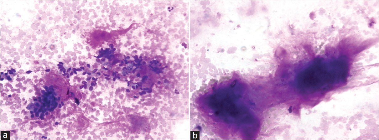
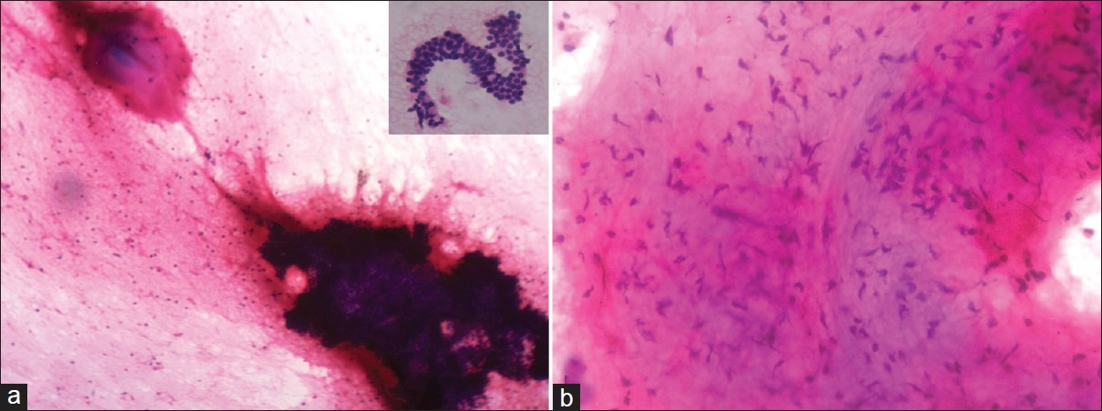
Fine needle aspiration cytology was done with 22 G needles. Two-to-three passes of the needle yielded a thick and mucoid aspirate. The smears were prepared and stained with May-Grunwald–Giemsa (MGG) and Haematoxylin-Eosin stains. The cellular smears showed a biphasic component-cellular and stromal elements. The cellular elements were composed of bland epithelial cells embedded in a fibrillary chondromyxoid stromal matrix [Figure 2]. The epithelial cells were small and monomorphic, with round-to-oval, centrally located nuclei, evenly dispersed fine chromatin and a moderate amount of cytoplasm. The cells were arranged in sheets and loose clusters with a few single cells [Figure 3a]. The elongated myoepithelial cells were embedded in a chondromyxoid matrix [Figure 3b]. A diagnosis of CS was made and a histopathological examination was advised.

- (a) Cellular smear shows relatively small, bland, monomorphic epithelial cells embedded in the myxoid ground substance (MGG, × 40); (b) The MGG stain highlights the fibrillary nature of the metachromatic chondromyxoid ground substance (MGG, × 40)

- (a) Cellular aspirates composed of bland, monomorphic cells arranged in sheets and loose clusters with few single cells in the myxoid background (Hematoxylin–Eosin stain, × 10). Inset shows groups of monomorphic epithelial cells having round-to-oval, centrally located nuclei, evenly dispersed fine chromatin and moderate amount of cytoplasm (Hematoxylin–Eosin stain, × 40); (b) Myoepithelial cells embedded in a chondromyxoid matrix (Hematoxylin–Eosin stain, × 40)
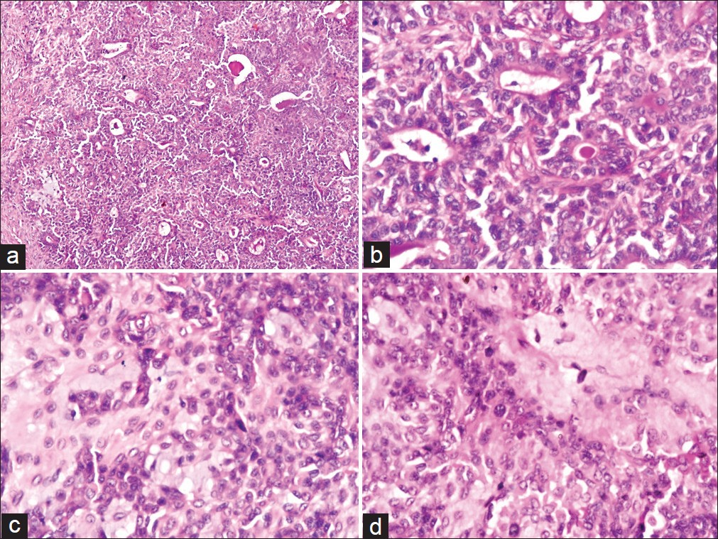
Under local anaesthesia, total excision of the lesion was performed, with disease-free margins. Grossly, the relatively well-circumscribed, small, epidermal tumour measured 0.9 cm in diameter. Microscopy revealed two components of the tissue. One component was made up of epithelial cells arranged in tubules, ducts and nests [Figure 4a]. They formed interconnecting tubuloalveolar structures lined by a single layer of cuboidal epithelial cells [Figure 4b]. The other component was made up of chondromyxoid stroma, intermingled with nests and branches of cuboidal epithelial cells [Figure 4c–d]. The histological findings were consistent with the benign mixed tumour of the skin - eccrine variant of chondroid syringoma. The patient was well after excision and no recurrence was reported on a two-year follow-up.

- (a) The histological section shows epithelial cells arranged in tubules, ducts and nests. (Hematoxylin–Eosin,× 10); (b) Interconnecting tubuloalveolar structures lined by a single layer of cuboidal epithelial cells (Hematoxylin–Eosin,× 40); (c,d) and chondromyxoid stroma, with nests and branches of cuboidal epithelial cells (Hematoxylin-eosin,× 40)
The aetiopathogenesis of CS is unknown.[4] The clinical characteristic of CS is not diagnostic.[1247] CS are commonly present as slowly growing, painless, firm, subcutaneous or intra-dermal nodules, measuring 0.5-3 cm in diameter, in the head and neck region of middle- and older-aged patients, with male predilection.[12467] The common sites are cheek, nose, or the skin above the lip.[1247] It is often single, but multiple CS have also been reported.[4] CS are often mistaken for other types of skin lesions, such as, dermoid or sebaceous cyst, neurofibroma, dermatofibroma, lipoma, basal cell carcinoma, pilomatricoma, histiocytoma and seborrheic keratosis.[46]
As CS are uncommon and rarely undergo aspiration, preoperative diagnoses of many cases are incorrect.[26] Very few cases describing the cytological features are available in the literature.[14] If the typical biphasic epithelial and mesenchymal elements are not represented on the aspirate, with one component predominating, it may lead to diagnostic difficulties and misdiagnoses.[1]
Fine Needle Aspiration Cytology is useful to determine benign and malignant CS before excision; however, sometimes it is difficult, because of the overlapping cytological features.[4] Examination of the excised tissue is most reliable in establishing a definitive diagnosis.[12] Histologically two variants are described by Headington, the eccrine type, with smaller lumens lined by a single row of cuboidal epithelial cells and the apocrine type, with tubular and cystic branching lumina lined by two rows of epithelial cells.[246] The inner layer expresses epithelial markers such as cytokeratin. The outer layers express mesenchymal markers such as vimentin, S-100 protein, neuron-specific enolase (NSE) and glial fibrillary acidic protein (GFAP).[127] CS is cytologically and histologically analogous with mixed tumours of the salivary gland. Even as pleomorphic adenomas arise from salivary glands, CS arises from sweat glands. The diagnosis of CS is dependent on the exact site of the lesion.[124]
Malignant CS is extremely rare.[14] Most malignancies develop de novo; few cases arise in the pre-existing benign CS.[18] Malignancy occurs more commonly in younger females, with a locally invasive lesion larger than 3 cm, involving the trunk and extremities.[45] Lymph node metastases and distant metastases are reported in 48 and 45 per cent of the cases, respectively.[4]
If a skin adnexal tumour is suspected clinically, the usual approach is biopsy, due to easy accessibility.[1] However, biopsy of CS has a risk of recurrence and malignant transformation.[12] Pre-operative cytological diagnosis of CS guides in the optimal management of the form of total excision with wide disease-free margins, without destroying the aesthetic and functional structures.[145] Regular follow-up is required to check for recurrence and malignancy.[1246]
To conclude, CS is often overlooked because of its rarity and unremarkable clinical presentation. CS is rarely aspirated. Awareness of the cytological features of CS is required in order to provide correct diagnosis and management. CS must be considered in the differential diagnosis of any small subcutaneous nodule in the head and neck region, in middle-aged patients. The treatment of choice is total excision, with wide disease-free margins, to rule out malignancy and reduce the risk of recurrence and malignancy in future.
REFERENCES
- Cytologic features of chondroid syringoma in fine needle aspiration biopsies: A report of 3 cases. Acta Cytol. 2010;54:183-6.
- [Google Scholar]
- Chondroid syringoma Mixed tumour of skin, salivary gland type. Arch Dermatol. 1961;84:835-47.
- [Google Scholar]
- Fine needle aspiration cytology of chondroid syringoma. Coll Antropol. 2010;34:687-90.
- [Google Scholar]
- Chondroid syringoma: A diagnosis more frequent than expected. Dermatol Surg. 2003;29:179-81.
- [Google Scholar]
- Fine needle aspiration cytology of malignant chondroid syringoma: A case report. Acta Cytol. 1998;42:1155-8.
- [Google Scholar]


