
Translate this page into:
Expanding the dermatological acronym from BLEND TAN EGG to PEGG: A rare case of painful palmar neurofibroma

*Corresponding author: Vinolyn D’Souza, Department of Dermatology Venereology and Leprosy, Father Muller Medical College, Mangaluru, Karnataka, India. vinolyndsouza@gmail.com
-
Received: ,
Accepted: ,
How to cite this article: D’Souza V, Bhat RM, D’Souza MJ, Fernandes MS. Expanding the dermatological acronym from BLEND TAN EGG to PEGG: A rare case of painful palmar neurofibroma. J Cutan Aesthet Surg. doi: 10.25259/JCAS_28_2024
Abstract
Painful skin tumors present a diagnostic challenge due to their diverse presentations. The acronym blue rubber bleb nevus, leiomyoma, eccrine spiradenoma, neuroma, dermatofibroma, tufted angioma, angiolipoma, neurilemmoma, endometrioma, granular cell tumor, and glomus tumor (BLEND TAN EGG) encompasses some of the common causes of painful skin tumors. This case report documents a 24-year-old woman presenting with a painful, solitary palmar nodule confirmed as a neurofibroma after an excisional biopsy. Neurofibromas are frequently asymptomatic, but their location within the tight palmar fascia can lead to significant discomfort. The rarity of this presentation and the difficulty in pre-operative diagnosis highlight the importance of considering palmar neurofibromas as a differential diagnosis for painful skin tumors, particularly in the hand and feet. To enhance the educational value of the existing acronym, we propose its expansion to “BLEND TAN PEGG,” where “P” signifies palmar or plantar neurofibroma.
Keywords
Painful skin tumors
Palmar neurofibroma
Plantar neurofibroma

INTRODUCTION
Peripheral nerve tumors constitute <5% of all hand tumors.1 They are classified as benign or malignant. Fortunately, the vast majority of them occurring on the hand are benign, with the most commonly encountered types being schwannomas and neurofibromas.
Neurofibromas are non-encapsulated tumors derived from Schwann cells and can be solitary or multiple. Multiple lesions may indicate von Recklinghausen’s disease, necessitating evaluation for systemic involvement. The solitary lesions are more common, and their growth in confined spaces like the hand can lead to discomfort or pain. In many instances, the diagnosis can only be made by excisional biopsy.2,3 Our case represents such a lesion and, hence, needs to be considered as a differential when encountering painful skin tumors in clinical practice.
Acronyms, created from the initial letters of words, serve as vital educational tools for recalling complex medical information. This case report proposes the extension of the existing acronym for painful skin tumors.4
CASE REPORT
A 24-year-old woman presented with a nodule on her right palm, gradually enlarging over 2 years. The nodule, initially asymptomatic, had started to cause significant pain, disrupting her daily activities over the past month. There was no noted history of trauma, no similar lesions elsewhere, and her family history was non-contributory.
On examination, a solitary skin-colored, smooth-surfaced nodule approximately 2 × 1 cm was present over the hypothenar aspect of her right palm. It was firm, fixed, and tender on palpation with a Wong-Baker Faces pain rating scale score of 6 [Figure 1].
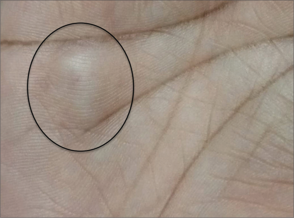
- Well-defined, skin-colored, and solitary nodule over the hypothenar aspect of the right hand (encircled).
Keeping the differentials of painful tumors in mind, ultrasonographic evaluation characterized the nodule as a well-defined hypoechoic mass within the subcutaneous tissue. Fine-needle aspiration yielded scant cellular material and mature adipose tissue stromal fragments in a background of hemorrhage and fat droplets suggestive of a lipomatous lesion. For a definitive diagnosis, an excision biopsy was undertaken. Histopathological analysis revealed a tumor composed of spindle cells arranged in fascicles. Individual cells possessed buckled nuclei, inconspicuous nucleoli, and a dense eosinophilic cytoplasm, features consistent with a neurofibroma [Figure 2]. There was no evidence of atypia. Immunohistochemical staining was positive for S100 protein [Figure 3], reinforcing the histopathological impression.
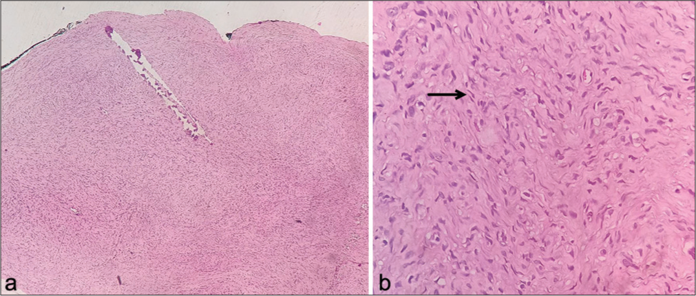
- (a) Tumor composed of spindle cells arranged in fascicles. (b) Plenty of individual cells have buckled nuclei, inconspicuous nucleoli, and eosinophilic cytoplasm, some resembling “diving dolphins” (arrow), embedded in a mucopolysaccharide stroma. Hematoxylin and eosin, ×40 (a) and ×400 (b).
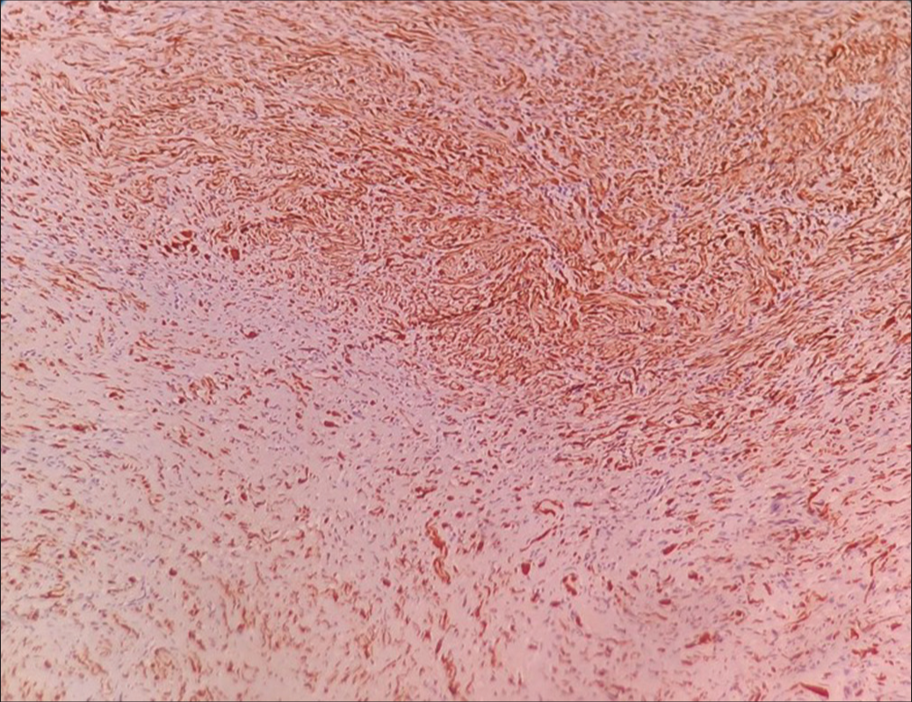
- Neural cells showing positive S100 immunoreactivity.
In the context of a solitary neurofibroma, a thorough systemic workup, including a fundoscopic examination and neuroimaging, was essential to rule out the possibility of von Recklinghausen’s disease. These investigations did not uncover any additional abnormalities. Furthermore, the family history did not reveal any evidence of consanguinity. As solitary neurofibromas are predominantly sporadic, mutational analysis was not pursued.
Thus, the lesion was diagnosed as an isolated acquired palmar neurofibroma.
DISCUSSION
Neurofibromas are World Health Organization grade 1 benign peripheral nerve sheath tumors consisting of diffuse proliferation of cells of neural and mesenchymal origin. Neurofibromas can be classified as localized (the most frequent), diffuse, or plexiform. Most cases are solitary and sporadic (90%), and the rest occur in the setting of neurofibromatosis type 1 (NF1) (10%), where they are usually multiple. The localized form of neurofibromas has an extremely low risk of malignant transformation.1
Neurofibromas are caused by NF1 gene deletions in both sporadic and syndromic cases. In sporadic cases, only the lesional cells exhibit this mutation, as compared to the germline mutation in NF1.5,6
Localized neurofibromas may manifest across various body parts, predominantly on the trunk, head/neck, and extremities, where they typically present without symptoms. These tumors are considered relatively uncommon in hand, accounting for <5% of all tumors found in this region; however, when they do appear in this area, particularly near the flexion creases, they may lead to discomfort and nerve-related issues due to the tight, rigid fascia of the glabrous palm.2,7
Clinically, these hand masses may often appear as a soft flesh-colored papule or small subcutaneous nodule and are histologically characterized by low cellularity of spindle cells with poorly defined cell borders in a myxoid to a pale pink collagenous matrix. The nuclei of these cells may be small, hyperchromatic, and wavy, giving them a “buckled” or “comma-shape,” resembling “diving dolphins.” These tumors are S-100 positive and often contain mast cells.8
Neurofibromas tend to involve the central portion of the nerve and blend with surrounding tissues, making it very difficult to enucleate the tumor without damaging the nerve fibers.3 In a study conducted by Lincoski et al. on benign tumors of the hand,2 the correct pre-operative diagnosis was made in only 1 (4.2%) of the 24 cases, indicating the need for strong clinical suspicion when clinicians encounter painful skin tumors in day-to-day practice. Only then can it be carefully dissected from the involved nerve under magnification, and nerve function can be preserved.
On axial T2W magnetic resonance imaging, a central hypointensity surrounded by a hyperintense rim, known as the target sign, is most commonly associated with neurofibromas, but it lacks both sensitivity and specificity for definitive diagnosis.9
Surgery is considered necessary for lesions over 4 cm or suspected neurofibromas that cause pain, numbness, or reduced range of motion.1,3 Despite their non-encapsulated nature and tendency to blend with nerve fibers, our case demonstrated successful excision without nerve damage.
The literature suggests a generally low risk of recurrence for solitary, sporadic neurofibromas when completely excised with clear margins. As in our case, there was successful excision of the neurofibroma without any evidence of atypia in the histological examination. This lowers the chance of regrowth. However, it is important to note: Long-term follow-up data on solitary palmar neurofibromas are limited.1
In our exploration of painful tumors of the skin, the acronym BLEND TAN EGG- blue rubber bleb nevus, leiomyoma, eccrine spiradenoma, neuroma, dermatofibroma, tufted angioma, angiolipoma, neurilemmoma, endometrioma, granular cell tumor, and glomus tumor has proven to be an invaluable educational tool.4
Our case, presenting a rare instance of a palmar neurofibroma, compels us to propose an addition to this acronym: the letter “P” to create BLEND TAN PEGG.
This expansion serves to refine the mnemonic as a more comprehensive educational aid.
CONCLUSION
This rare case highlights the importance of considering palmar neurofibromas in the differential diagnosis of painful skin tumors. The proposed expansion of the acronym from BLEND TAN EGG to PEGG by incorporating palmar or plantar neurofibromas aims to aid clinicians in recalling a broader spectrum of differential diagnoses for painful skin tumors, ultimately enhancing diagnostic accuracy and patient management.
Authors’ contributions
Dr(s) Vinolyn D’Souza, Ramesha Bhat M, Myfanwy Joanne D’Souza, and Michelle Serene Fernandes had full access to the study data and were responsible for ensuring its integrity and accuracy. All authors were involved in editing and reviewing the manuscript, in addition to their specific contributions listed below: Vinolyn D’Souza: Conceptualization, study design, intellectual content definition, literature search, data acquisition, and manuscript preparation. Ramesha Bhat M: Conceptualization, study design, and intellectual content definition. Myfanwy Joanne D’Souza: Study design, literature search, and manuscript preparation. Michelle Serene Fernandes: Manuscript preparation and critical revision.
Ethical approval
The case report was approved by Father Muller Institutional Ethics Committee, number 035/2024, dated January 01, 2024.
Declaration of patient consent
The authors certify that they have obtained all appropriate patient consent.
Conflicts of interest
There are no conflicts of interest.
Use of artificial intelligence (AI)-assisted technology for manuscript preparation
The authors confirm that there was no use of artificial intelligence (AI)-assisted technology for assisting in the writing or editing of the manuscript, and no images were manipulated using AI.
Financial support and sponsorship
Nil.
References
- Peripheral nerve tumors of the hand: Clinical features, diagnosis, and treatment. World J Clin Cases. 2020;8:5086-98.
- [CrossRef] [Google Scholar]
- Benign nerve tumors of the hand and the forearm. Am J Orthop (Belle Mead NJ). 2007;36:E32-6.
- [Google Scholar]
- Tumours of peripheral nerves in the upper extremity: A 22-year epidemiological study. Scand J Plast Reconstr Surg Hand Surg. 2009;43:43-9.
- [CrossRef] [Google Scholar]
- Painful tumors of the skin-from ENGLAND to LEND AN EGG to BLEND TAN EGG. Indian J Dermatol Venereol Leprol. 2019;85:231-4.
- [CrossRef] [Google Scholar]
- S100B and neurofibromin immunostaining and X-inactivation patterns of laser-microdissected cells indicate a multicellular origin of some NF1-associated neurofibromas. J Neurosci Res. 2011;89:1451-60.
- [CrossRef] [Google Scholar]
- Solitary neurofibromas: Does an uncommon site exist? Ann Dermatol. 2012;24:101-2.
- [CrossRef] [Google Scholar]
- Practical approach to histological diagnosis of peripheral nerve sheath tumors: An update. Diagnostics (Basel). 2022;12:1463.
- [CrossRef] [Google Scholar]
- Telltale signs of peripheral neurogenic tumors on magnetic resonance imaging. Indian J Radiol Imaging. 2015;25:453-8.
- [CrossRef] [Google Scholar]


