Translate this page into:
Extradiagnostic Use of Dermoscopy: A Report of Two Cases
Address for correspondence: Yasmeen Jabeen Bhat, Department of Dermatology, Venereology and Leprosy, GMC Srinagar, Jammu and Kashmir, India. E-mail: yasmeenasif76@gmail.com
This article was originally published by Wolters Kluwer - Medknow and was migrated to Scientific Scholar after the change of Publisher.
Abstract
Abstract
Dermoscopy refers to evaluation of the skin surface using surface microscopy. It is mainly used for the diagnosis of skin disorders. We report two cases in which dermoscopy played a role in treatment. Our first case was a 40-year-old female with history of insect bite. We evaluated the patient using a dermoscope and removed the tick with mouth part embedded in dermis using forceps ensuring full removal after procedure. The second case was a 35-year-old female who presented with a non-healing ulcer over lower back, following excision of epidermoid cyst. Dermoscopy showed the presence of a thread which was removed and repeat dermoscopy following extraction ensured its full removal.
Keywords
Dermoscopy
extradiagnostic use
foreign body
insect bite
INTRODUCTION
Dermoscopy, also referred to as dermatoscopy or epiluminescence microscopy, is used for the evaluation of skin surface using surface microscopy. It was initially used for diagnosis of melanomas and non-melanoma skin cancers. Dermoscopy now finds its use in the diagnosis of a wide range of skin conditions such as inflammatory disorders, connective tissue disorders, infections, and so on.[1] The use of dermoscopy has come a long way and in recent years its use has been extended for the treatment of a variety of conditions. Some of the extradiagnostic uses of dermoscopy include disease activity evaluation, ex-vivo histological evaluation, pre- and post-treatment comparison, and evaluation of therapeutic modalities in clinical studies. It also improves doctor–patient communication.[2]
Detection and removal of foreign bodies and retained sutures, for example, black threads, especially in dark skin types, can be difficult due to lack of color contrast between object and skin.[3] In these cases, dermoscopy can be of great help by providing a three-dimensional view that enables proper detection and removal of these substances.[4]
We report two cases in which dermoscopy helped in treatment. adding to extradiagnostic use of dermoscopy.
CASE 1
Our first case was a 40-year-old female who presented to us with chief complaints of insect bite over right flank for the past 5 days. The lesion was associated with mild-to-moderate itching. On examination, we noted a brown-colored papule of size 4 × 3 mm with surrounding red-colored macule of 4 × 3 cm [Figure 1]. Patient gave history of relief in pruritus from use of over-the-counter antihistamines. Dermoscopy of the lesion showed the presence of a black grayish surface of the tick with eight legs on the sides and mouth piece embedded in the skin [Figure 2]. We removed the tick using forceps, viewing through the dermoscope, and dermoscopy after procedure ensured its full removal with visualization of hemorrhages only [Figure 3]. Ex vivo dermoscopy showed the tick with intact mouth piece on a brownish white scale [Figure 4].
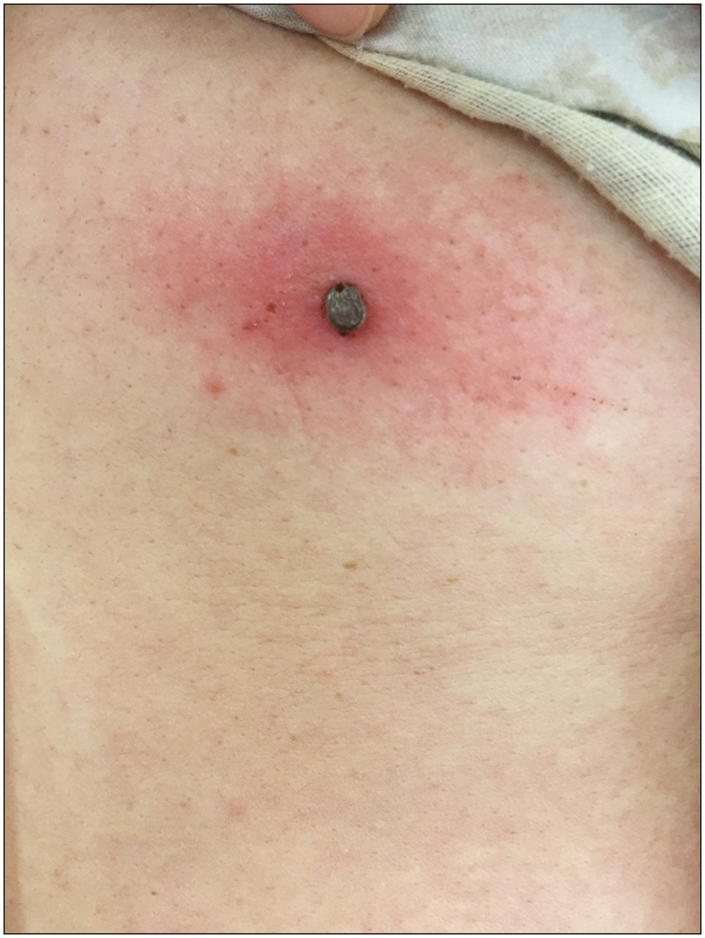
- Clinical image of insect bite on the back
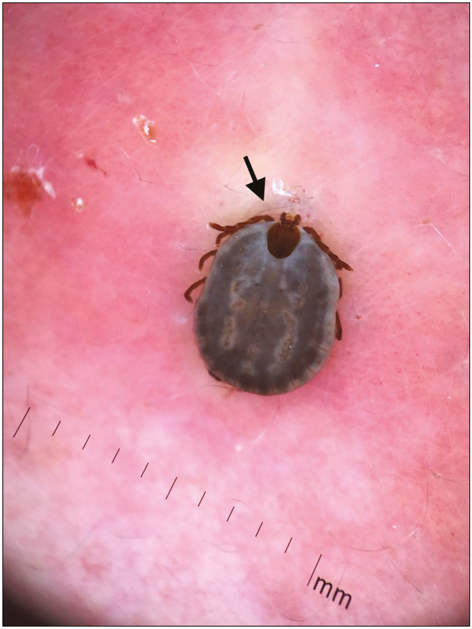
- Dermoscopy showing the blackish gray back of the tick with four pairs of dark brown legs on either side and dark mouth piece (black arrow) (polarized mode, DermLite DL3, 10×)
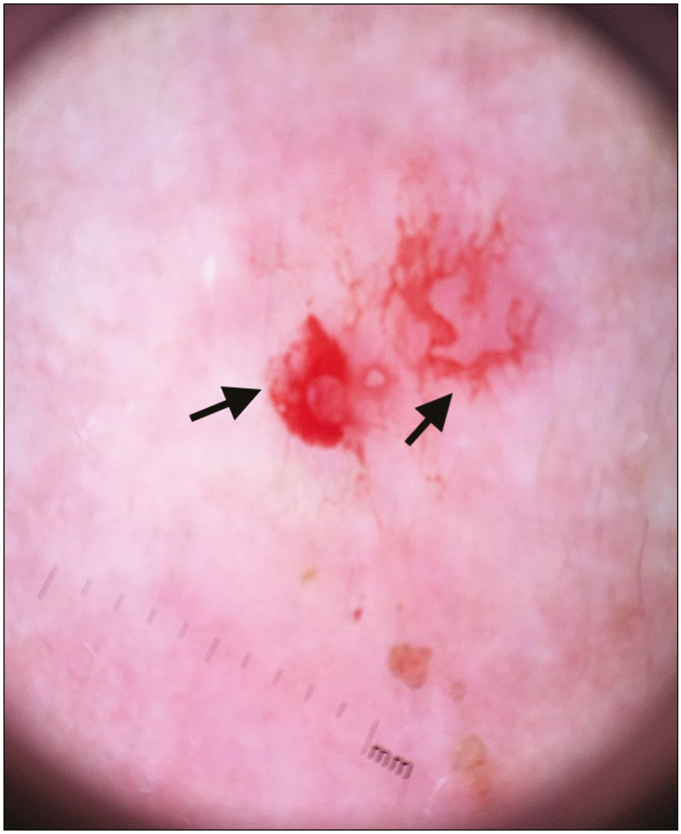
- Dermoscopy revealing blood spots and hemorrhagic areas (black arrows) with no remnants after removal of the tick (polarized mode, DermLite DL3, 10×)
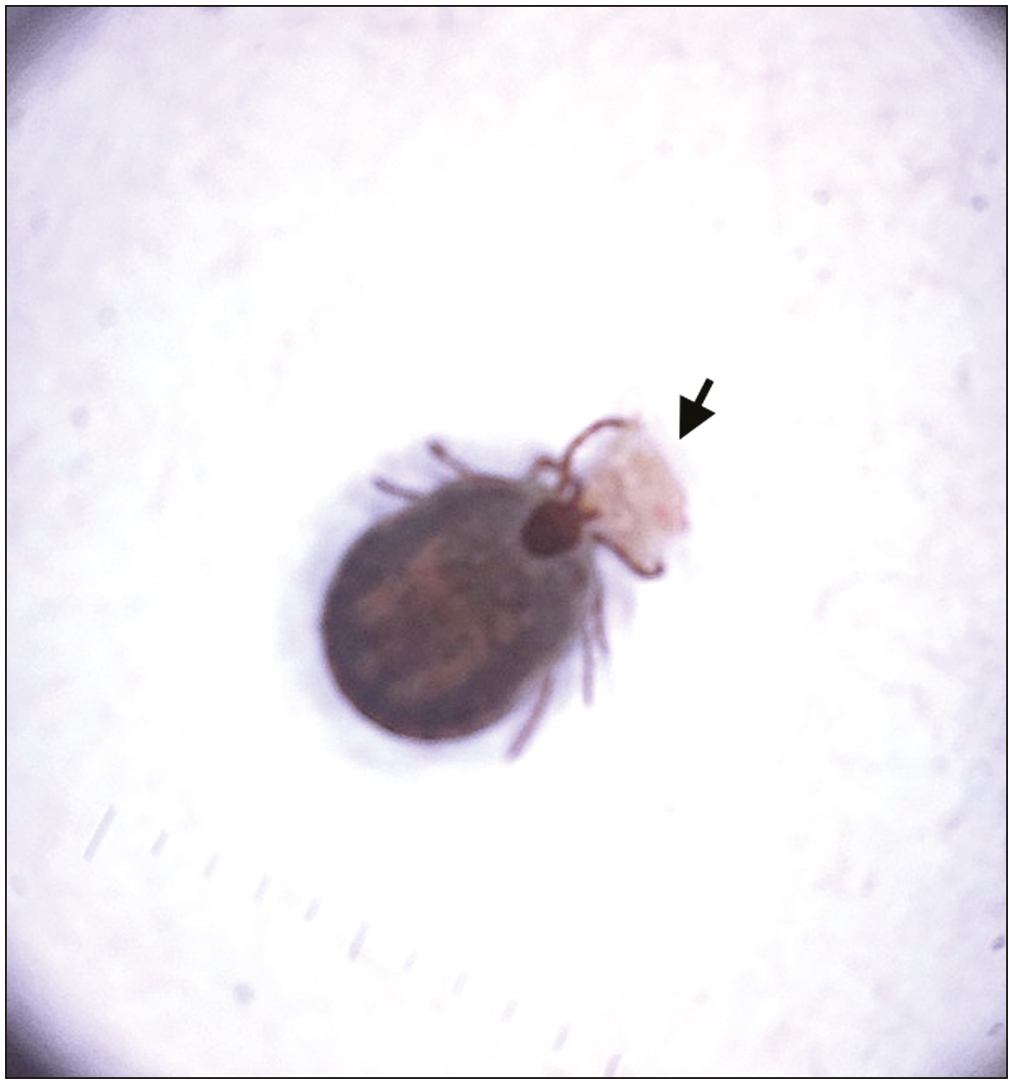
-
Ex-vivo dermoscopy showing the tick with intact mouth piece on a brownish white scale (black arrow) (polarized mode, DermLite DL3, 10×)
CASE 2
Our second case was a 35-year-old female who presented to us with chief complaints of non-healing ulcer over lower back since few weeks, not responding to antibiotics. The patient gave history of excision of biopsy-documented epidermoid cyst over the same site a month back. Other history was unremarkable, and on examination, we found a violaceous plaque of size 1.5 × 1 cm with overlying yellow crust in the center [Figure 5]. The lesion was tender on palpation and base was non-indurated. Dermoscopy of the lesion showed the presence of a black-colored thread inside the yellowish brown homogeneous area [Figure 6]. It was removed using forceps and dermoscopy after procedure ensured full removal of the suture. The patient was prescribed topical and systemic antibiotics and was lost to follow-up.
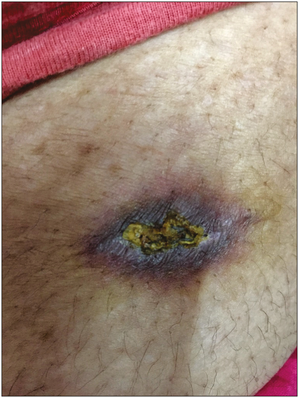
- Clinical image of non-healing ulcer on the back
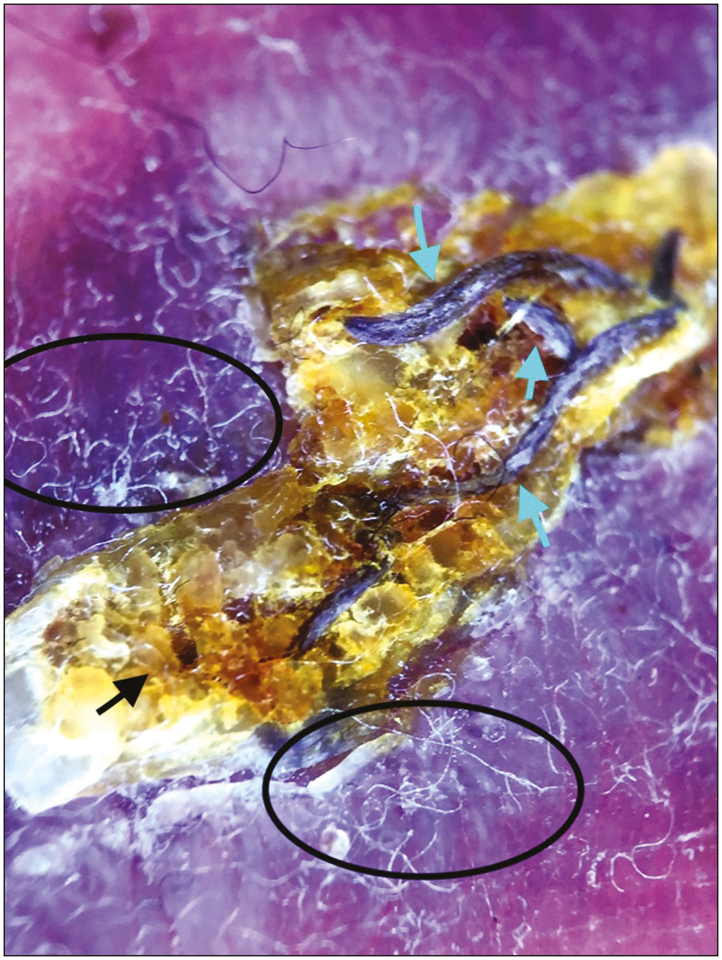
- Dermoscopy showing blackish suture material (green arrows) embedded in the brownish yellow homogeneous crust (black arrow), with surrounding adherent fibers (black circles) (polarized mode, DermLite DL3, 10×)
DISCUSSION
Dermoscopy is emerging as a tool, playing role in the treatment of various skin disorders. The use of dermoscopy for evaluation of skin infestations is referred to as entomodermoscopy. It finds its use in diagnosis and management of infestations such as scabies, pediculosis, myiasis, tick bites, and so on. Ticks are blood sucking arthropods which attach to the human skin by specialized mouth pieces. After biting, the mouth pieces of ticks remain embedded in the human skin and can lead to persistent reaction in the immediate phase and formation of foreign body granuloma in the late phase.[5] In our case, dermoscopy enabled removal of the mouth piece under magnification, thus ensuring full removal.
Dermoscopy has been used for detection and extraction of intracutaneous foreign bodies. In our case, we were able to diagnose the cause of non-healing ulcer using dermoscopy as well as treatment of the same by extracting the thread.
These two cases provide evidence showing expanding applications of dermoscopy beyond its diagnostic use.
Declaration of patient consent
The authors certify that they have obtained all appropriate patient consent forms. In the form the patient(s) has/have given his/her/their consent for his/her/their images and other clinical information to be reported in the journal. The patients understand that their names and initials will not be published and due efforts will be made to conceal their identity, but anonymity cannot be guaranteed.
Financial support and sponsorship
Nil.
Conflicts of interest
There are no conflicts of interest.
REFERENCES
- Dermoscopy in general dermatology: A practical overview. Dermatol Ther (Heidelb). 2016;6:471-507.
- [Google Scholar]
- Dermoscopy Overview and Extradiagnostic Applications. Treasure Island, FL: Stat Pearls Publishing; 2022.
- Dermoscopy update: Review of its extradiagnostic and expanding indications and future prospects. Dermatol Pract Concept. 2019;9:253-64.
- [Google Scholar]
- Monitoring of therapeutic response and other applications. In: Lallas A, Errichetti E, Ioannides D, eds. Dermoscopy in General Dermatology. Boca Raton, FL: CRC Press; 2018. p. :325-9.
- [Google Scholar]


