
Translate this page into:
Innovative use of needles, connectors, and syringes in follicular unit extraction
*Corresponding author: K. Shilpa, Department of Dermatology, Bangalore Medical College, Bangaluru, Karnataka, India. shilpakvinod1980@gmail.com
-
Received: ,
Accepted: ,
How to cite this article: Narayan VR, Shilpa K, Eswari L. Innovative use of needles, connectors, and syringes in follicular unit extraction. J Cutan Aesthet Surg. doi: 10.25259/JCAS_80_2024
Abstract
Follicular unit extraction (FUE) is an effective but physically demanding hair transplantation technique. Operator fatigue and technical challenges, such as slit dilation, graft maintenance, and tumescent anesthesia administration, can complicate the procedure. This study introduces cost-effective, innovative solutions using commonly available operating theater tools – needles, connectors, and syringes – to address these challenges. A modified hypodermic needle bent at acute angles is proposed to aid in slit dilation, improving graft placement accuracy and preventing graft dislodgement, especially in confined areas between implanted follicles. In addition, a three-way connector attached to a syringe and IV drip set provides continuous irrigation, ensuring a bloodless field and maintaining graft moisture without manual compression or refilling. Finally, a three-way connector is used to streamline the administration of tumescent anesthesia, minimizing the risk of needle stick injuries while maintaining sterility. These modifications simplify the FUE procedure, enhance maneuverability, and reduce surgeon fatigue, contributing to improved surgical outcomes. By repurposing everyday tools, this technique offers an accessible and effective means of overcoming common challenges in FUE, potentially revolutionizing standard hair transplantation practices.
Keywords
Follicular unit extraction
Hypodermic needle
Surgical pearl

PROBLEM STATEMENT
FUE presents clinical challenges, particularly in the identification and dilation of slits, especially when recipient sites are narrower than the implanted grafts. Operator fatigue, uncontrolled hand movements, and tremors further complicate the procedure. Traditional methods utilizing two forceps pose challenges in terms of dexterity, coordination, space utilization, bleeding, and visualization of grafts. This research proposes a simple modification to address these challenges.
RECOMMENDED SOLUTION
We introduce a novel technique employing a modified hypodermic needle bent at acute angles to act as a slit dilator [Figure 1]. This modification facilitates the gentle lifting and spreading of slits, enhancing visualization and maneuverability. The bend in the needle proves advantageous, especially in grafting areas between implanted follicles, preventing graft pop-out. Furthermore, the technique prevents overcrowding and allows multiple implanters to work simultaneously, as demonstrated in Video 1. Notably, no additional equipment is required, as the same needle used for infiltrating tumescent anesthesia can be repurposed for this modified procedure. The needles can be bent to different angles to help implantation in different parts of the scalp. More acute angle would be used for the temporal region. The frontal region can have a bend of 30° and the parietal region will have a bend of 45°.
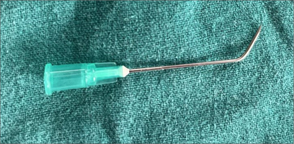
- The modification involving a 20 G hypodermic needle bent to craft an improvised slit dilator. This bent design enhances slit visualization while the tip effectively engages with the slits during the procedure.
Video 1:
Video 1:Showcasing the practical application of the modified hypodermic needle, enabling precise implantation with enhanced dexterity. The effortless dilation not only supports atraumatic graft implantation but also contributes to a reduced learning curve for practitioners.PROBLEM STATEMENT
Maintaining graft moisture and a bloodless field during FUE implantation requires continuous irrigation. Conventionally, this is achieved using a spray canister or a syringe with a broken needle. However, using a syringe and broken needle necessitates frequent refilling, while the spray canister may not generate enough force to clear blood and clots and requires manual compression each time, leading to surgeon fatigue during lengthy procedures such as hair transplantation.
RECOMMENDED SOLUTION
An improvised device can be created by attaching a broken needle to a syringe through a three-way connector. The other opening of the connector is connected to an IV drip set attached to a saline bag. The broken needle port and drip set ports are made active by turning the three-way knob. The drip set is allowed to flow at maximal rate. The IV drip set runs freely, allowing continuous, gentle irrigation of the field. If more force is needed, the connector can be turned to connect the IV drip to the syringe. Withdrawing the plunger fills the syringe, and turning the connector to link the broken needle to the filled syringe allows for greater irrigation force to dislodge clots if present [Videos 2 and 3].
Video 2:
Video 2:Showcasing the interrupted spray and continuous spray modes of the three-way connector innovation. This improves the efficacy of the technique and ensures the grafts are hydrated.Video 3:
Video 3:Demonstration of the working of the syringe connector innovation using methylene blue mixed with saline solution as irrigating fluid.PROBLEM STATEMENT
Hair transplantation requires significant amounts of tumescent anesthesia, whether during slit making or FUE. Reloading the syringe with the tumescent solution can be time-consuming, and the needle may become blocked. Frequent removal and reattachment of the needle can lead to needle stick injuries and compromise sterility.
RECOMMENDED SOLUTION
A three-way connector can be used to attach the needle to one port, a syringe to another, and a drip set connected to a tumescent solution bag to the third. By allowing the drip set to flow freely and turning the connector so that the drip set and syringe ports are active, the syringe can be filled with a tumescent solution by withdrawing the plunger. The connector is then turned to activate the syringe and needle ports. The needle is inserted into the scalp, the plunger is withdrawn to ensure the needle is not intravascular, and then the plunger is pushed to deliver the tumescent solution into the target area. This method allows for the reloading of the syringe without removing the needle, maintaining sterility and reducing the risk of needle stick injuries [Video 4].
Video 4:
Video 4:Showcasing the technique of tumescence solution delivery and refilling using the three-way connector. This reduces operator fatigue and improves asepsis.Author contributions
R. Vignesh Narayan: conceptualization, methodology, writing – original draft. K. Shilpa: data collection, formal analysis, writing – review and editing, supervision, project administration. L. Eswari: investigation, resources, writing – review and editing.
Ethical approval
Institutional Review Board approval is not required.
Declaration of patient consent
The authors certify that they have obtained all appropriate patient consent.
Conflicts of interest
There are no conflicts of interest.
Use of artificial intelligence (AI)-assisted technology for manuscript preparation
The authors confirm that there was no use of artificial intelligence (AI)-assisted technology for assisting in the writing or editing of the manuscript and no images were manipulated using AI.
Financial support and sponsorship: Nil.


