Translate this page into:
Insects are Crawling in My Genital Warts
Address for correspondence: Dr. Somesh Gupta, Department of Dermatology and Venereology, All India Institute of Medical Sciences, New Delhi 110 029, India. E-mail: someshgupta@hotmail.com
This is an open-access article distributed under the terms of the Creative Commons Attribution-Noncommercial-Share Alike 3.0 Unported, which permits unrestricted use, distribution, and reproduction in any medium, provided the original work is properly cited.
This article was originally published by Medknow Publications and was migrated to Scientific Scholar after the change of Publisher.
Abstract
A 23-year-old woman presented with large exophytic genital wart arising from perineum, vulva, introitus of the vagina, and inner aspect of thighs. Patient developed severe itching and formication (insect-crawling sensation) in the lesions for past 1 week, though careful examination did not reveal any insects. Considering that the disease was causing psychological stress and physical symptoms, radiofrequency excision was planned. However, during the procedure, several maggots appeared from the crypts. The procedure was abandoned and maggots were removed manually. Subsequently external giant warts were removed using radiofrequency device. There was no recurrence of excised warts during 3 month follow-up. To our knowledge, this is the second reported case of maggots in genital warts.
Keywords
Genital warts
myiasis
radiofrequency excision
INTRODUCTION
Psychiatric and psychological morbidity has been reported in patients with genital warts.[1] Even though, genital warts are generally not associated with severe symptoms, they have profound adverse impact on quality of life of patients.[2] If patient with genital warts, complains of severe and unusual symptoms are often considered psychological. We report a case of giant genital warts where the patient complained of formication (insect-crawling sensation) which was subsequently found to be due to maggot infestation in the warts.
CASE REPORT
A 23-year-old married woman presented with large exophytic growth arising from perineum, vulva, introitus of the vagina, and inner aspect of thighs for past 4 months [Figure 1a]. There was a history of rapid growth in the lesions for past 2 months. Patient developed severe itching and formication in the lesions for 1 week, though she had never seen or recovered any insect. The large growth resulted in abnormal gait of the patient. Sexual history of the patient and her husband was unremarkable. In view of giant genital wart, serum enzyme-linked immuno sorbent assay (ELISA) for HIV and venereal disease research laboratory (VDRL) test for syphilis was done for both husband and the wife but were negative. Also, there was no other evidence suggestive of immunosuppression.

- (a) Giant genital wart before intervention. (b) Intraoperative picture showing maggot. (c) Three months after complete excision using a radiofrequency device
Cutaneous examination revealed an approximately 10 × 5 cm hyperpigmented, pedunculated growth with verrucous surface over both the labial folds symmetrically, completely obliterating the introitus. On per speculum examination, the verrucous lesions were found to involve the vaginal mucosa as well. Anal opening was also partially covered by the mass. Smaller verrucous papules and plaques were present in groin folds and perianal region. A diagnosis of giant condylomata acuminata was made and it was thought that patient's unusual complaint about the ‘crawling insects’ is related to the psychological stress caused by the disease. The HPV viral genotyping using linear array (Roche) showed HPV 6 and 11. Histopathology from the warts did not show any evidence of Bushke Löwenstein tumor or squamous cell carcinoma. On histopathological examination hyperkeratosis, papillomatosis, koilocytosis and dilated and congested capillaries in the dermis were found.
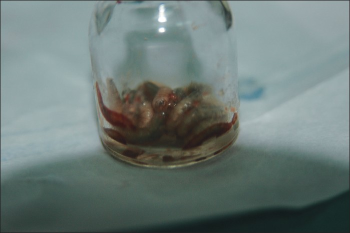
Within few days of presentation, she started complaining of paroxysms of severe perineal scratching consequent to increased sensation of crawling insects. But clinical examination again did not reveal any insects. Thinking that the disease was causing immense psychological stress hence leading to the strange symptoms, serial excision of the warts with a radiofrequency device (Ellman, Oceanside, NY, USA) was planned. During the procedure, periodic perineal spasms were observed. Local anaesthetic (2% lignocaine with adrenaline) was infiltrated in the lesion and the procedure was started. Suddenly, perhaps due to irritation caused by radiofrequency current, multiple live maggots were seen coming out of verrucous masses [Figure 1b]. The procedure was abandoned and thick white petrolatum was applied to the area. Approximately 20 live maggots were then removed with the help of forceps [Figure 2] and few of them were sent for entomological examination. The maggots were identified as those belonging to Chrysomia bezziana. The patient was given intravenous antibiotics (ceftriaxone and amikacin) in view of some denuded area produced due to radiofrequency excision of few warts. A magnetic resonance imaging (MRI) of abdomen and pelvis was done to rule out deeper soft tissue infestation and damage by the maggots which turned out to be normal. Patient's symptoms completely resolved with few more sessions of extraction of maggots over the next 2 days and subsequent examination did not reveal any maggots.

- Some of the removed maggots
Later, external genital warts were completely excised with radiofrequency [Figure 1c]. There was no recurrence of excised warts in 4-month post-operative follow-up. Warts inside the vagina are being treated with imiquimod and at the time of writing this report, they have reduced substantially in size and this treatment is being continued.
DISCUSSION
Myiasis is a parasitic infestation of human or animal skin, necrotic tissues and natural cavities by fly larvae or pupa. Cutaneous involvement is the most common type of myiasis. Human myiasis is caused by fly larvae capable of penetrating body orifices as well as healthy or necrotic tissue. This disease usually affects children and occurs predominantly in individuals with poor hygienic practices. Patton divided myiasis-causing flies into three parasitologic categories: obligatory, facultative, and accidental.[3] Our patient probably qualifies for facultative myiasis. Cochliomyia hominivorax, Chrysomya bezziana, and Wohlfahrtia magnifica are the most common flies worldwide that cause human wound myiasis.[4] Chrysomia bezziana is the most common cause of cutaneous myiasis in India but usually infests wounds and mucous membranes. Predisposing factors for human wound myiasis include open wounds, poor hygiene, advanced age, psychiatric illness, diabetes, vascular occlusive disease, and physical handicap.
The clinical manifestations of wound myiasis vary according to the affected body area and the extent of the infestation. Patients may present with fever, pain, bleeding from the infested site or may be complicated by secondary infection and tetanus. Genital/vulval myiasis is rare.[45] Genital condyloma with myiasis is even rarer. As far as ascertained, there is only one report in literature on myiasis in genital warts from Brazil in which the patient was pregnant and after removal of maggots the pregnancy ended in spontaneous abortion.[6] Myiasis in our case was unusual in view of its site and its occurrence in the absence of the usual risk factors. Due to rarity of condition, and our unfamiliarity we did not consider infestation by maggots though patient complained of crawling sensation and severe itching in the genital warts. The lesson learnt is to examine and investigate the patient thoroughly before dismissing patient's complaints as irrelevant, psychogenic or functional, however unusual and unexplainable they are for the disease.
The simplest treatment for myiasis is application of an occlusive agent such as white soft paraffin, wax, glue, adhesive tape or chewing gum followed by physical removal of maggots. Lidocaine or pilocarpine can be instilled to paralyze the worm to facilitate removal.[7]
Source of Support: Nil.
Conflict of Interest: None declared.
REFERENCES
- Major depressive episode secondary to condylomata acuminata. Gen Hosp Psychiatry. 2010;32(446):e3-5.
- [Google Scholar]
- Health related quality of life in Indian patients with three viral sexually transmitted infections - herpes simplex virus-2, genital human papilloma virus, and human immunodeficiency virus. Sex Transm Infect. 2011;87:216-20.
- [Google Scholar]


