Translate this page into:
Intralesional chemical cautery of papulonodular acne
*Corresponding author: Muhammed Mukhtar, Mukhtar Skin Centre, Katihar Medical College Road, Katihar, Bihar, India. drmmukhtar20@gmail.com
-
Received: ,
Accepted: ,
How to cite this article: Mukhtar M. Intralesional chemical cautery of papulonodular acne. J Cutan Aesthet Surg. 2024;17:252-3. doi: 10.4103/JCAS.JCAS_129_22
Dear Editor,
Papulonodular acne lesions are inflamed papules and nodules (follicular or mini pilosebaceous cyst). At times, they are resistant to treatment, which leads to scarring on the face. For better cosmetic results, the acne lesion should be drained out, and its wall should be damaged at an early stage. Intralesional steroids and drainage of the contents are helpful in the early resolution of the lesions.1,2 However, the recurrence is more frequent, and there is a chance of atrophic scars. Here, the authors describe a new technique to drain and cauterize the lesions for a longer-lasting solution and better cosmetic results.
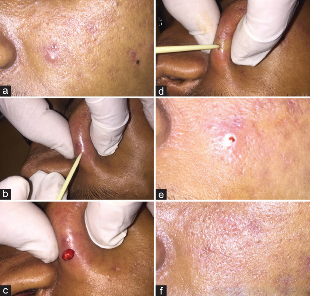
To drain and cauterize the papulonodular acne, the papule and nodule are pricked with a sterilized or disinfected toothpick, and the contents are removed manually [Figure 1a–c, Video 1]. Thereafter, the tip of the toothpick is dipped in trichloroacetic acid (TCA 100%) and reinserted into the drained wound to cauterize the wall of the lesion [Figure 1d and e, Video 2]. However, for cystic lesions, following incision and drainage, a cotton bud soaked in TCA or radiofrequency or hypertonic saline should be used to cauterize the cyst wall. After the procedure, the wounds are left open to heal. Following the procedure, patients are advised to apply topical antibiotics and moisturizers to keep the site infection free and moist for 7–10 days with general precaution for the wounds. These patients were followed weekly for 6–12 weeks. Frosting, burning sensation, moderate erythema and irritation shortly after cautery, and scabbing at the site for 5–10 days are common short-term consequences of this procedure. Wounds healed in 1–2 weeks with no recurrence of the treated lesions and long-term complications seen during follow-up [Figure 1f]. We have treated five patients with good cosmetic effects. However, to judge the effectiveness of the technique, case–control studies have to be conducted on a large number of patients.

- (a)-(c) nodule of acne on the face is pricked and drained; (d), (e) the drained acne lesions are intralesionally cauterized with trichloroacetic acid (TCA); (f) the acne nodule healed without scar.
Video 1:
Video 1:The nodulocystic acne lesions are pricked with toothpicks and drained.Video 2:
Video 2:The nodulocystic acne lesions are cauterized with TCA 100% using tooth prick.Authors’ contributions
Muhammed Mukhtar: Concepts, Design, Definition of intellectual content, Literature search, Manuscript preparation, Manuscript Editing, and Manuscript review.
Declaration of patient consent
The authors certify that they have obtained all appropriate patient consent forms. In the form the patient(s) has/have given his/her/their consent for his/her/their images and other clinical information to be reported in the journal. The patients understand that their names and initials will not be published and due efforts will be made to conceal their identity, but anonymity cannot be guaranteed.
Conflicts of interest
There are no conflicts of interest.
Videos available on:
Financial support and sponsorship
Nil.
References
- Innovative method for self-application of topical preparations on inaccessible sites. J Am Acad Dermatol. 2021;84:e225-6.
- [CrossRef] [PubMed] [Google Scholar]
- Surgical pearl: Disposable syringe as comedone extractor. Cosmetic Dermatol-Cedar Knolls. 2003;16:57-62.
- [Google Scholar]


