Translate this page into:
Pursestring Technique for Treatment of Nulliparous Congenital Inverted Nipple
Address for correspondence: Dr. Chetan Satish, Department of General Surgery, Sapthagiri Medical College, 67, 14th cross, 1st Block, R.T. Nagar, Bengaluru 560032, Karnataka, India. E-mail:drchetansatish5@gmail.com
This is an open access journal, and articles are distributed under the terms of the Creative Commons Attribution-NonCommercial-ShareAlike 4.0 License, which allows others to remix, tweak, and build upon the work non-commercially, as long as appropriate credit is given and the new creations are licensed under the identical terms.
This article was originally published by Wolters Kluwer - Medknow and was migrated to Scientific Scholar after the change of Publisher.
Abstract
Abstract
Background:
Nipple inversion in nulliparous women is often encountered and is a surgical challenge. The challenge lies in the correction of deformity with minimal marks and also in not damaging the lactiferous ducts, which will interfere with future breastfeeding.
Aim:
To provide a relatively simple technique, which can be easily performed as an outpatient procedure with minimal complications.
Materials and Methods:
A total of 11 patients were treated with this technique of which 5 patients had bilateral inverted nipples.
Results:
We had no recurrence and the follow-up period was of minimal of 1 year. One patient had mild stitch infection, which healed conservatively with antibiotics.
Conclusion:
The pursestring technique described offers a simple surgical option for the treatment of nulliparous women with nipple inversion.
Keywords
Inverted nipple
pursestring
ulliparous
INTRODUCTION
Nipple inversion is relatively common, occurring in up to 10% of females.[1]
Patients who present with inverted nipples typically are self-conscious and insecure about the appearance of their naked breasts. If the inverted nipple inhibits breastfeeding, it may impact parent–child bonding and impair the health of the child.[2]
Nipple inversion is caused either by failure of the lactiferous ducts to develop and grow during maturation of the breast tissue or by fibrosis around the lactiferous ducts due to inflammation (e.g., mastitis, cancer, previous breast surgery).[3] At approximately week 6 of fetal development, breast buds form along the milk line. The mammary glands develop as epithelial downgrowth into the mesenchymal tissue. Later, during the eighth or ninth month of fetal development, a pit forms at the entry to the ducts. The proliferation of mesenchymal tissue and fat below the pit causes it to elevate above the nascent skin to form the projection of the nipple. Failure of the growth of the mesenchyme or of lengthening of the lactiferous ducts can cause congenital inverted nipples.[4]
Inverted nipples can be classified into three categories based on the known classification systems and common findings.[3] Nipple inversion can be stratified according to the amount of eversion possible and the projection that can be maintained with traction. The surgical treatment of inverted nipples relies on several basic principles, including maintenance of as many of the functional lactiferous ducts as possible, meticulous dissection of the fibrous attachments surrounding the duct system, placement of mattress sutures at the nipple base to oppose the underlying tissue and close the dead space-maintaining eversion, placement of running sutures at the base of the nipple in selected cases, and stent utilization to maintain the nipple in a projected state as it heals.[3]
We have simplified the grades of nipple inversion for treatment as follows:
Grade 1: Proctactable easily without suction
Grade 2: Proctactable with suction
Grade 3: Not protractable with suction
MATERIALS AND METHODS
In our study, 11 patients were treated for congenital inverted nipples over a period from January 2015 to January 2018. Of these five patients had bilateral nipples. All patients were treated by similar technique with minimal follow-up of 1-year postop. None of patients had recurrence in follow-up period. No complications were reported except one patient who had mild stitch infection of external sutures which healed with antibiotics.
Surgical technique
All the cases were done under local anesthesia as an outpatient procedure using a magnifying loupe.
Basically the steps are as follows:
> Hold the nipple with toothed forceps.
> Take a traction stitch of 2-0 monofilament, with a good bite on nipple so it does not cut through.
> Take two diametrically opposite incisions, say 12 and 6 o’clock, at the junction of nipple and areola. Make them along the long axis of the nipple.
> Dissect the fibrous tissue by opening the blade of the scissors longitudinally along the direction of the ducts, not perpendicular to them. In nonlactating ducts, it is extremely difficult to see the ducts, even with magnification. Release as much as possible. The ductal structures were easily visualized and preserved during dissection. The ducts were visually identified as tubular nonvascular structures, larger and of different consistency than nerves. They did not resemble scar tissue.[5]
> Take a purse string with 3-0 monofilament material (prolene) around the base of the nipple and bring out through either incision. Tighten it without strangulating it.
> Close incisions with fine monofilament, and take an IV bottle cap, make 2 holes and tie the original 2-0 stay stitch using the cap as a bolster. Maintain traction for 3 to 4 days.
Once the pain has subsided and patient is comfortable, she should perform suction with the syringe three to four times daily. Previously we used a 20-mL Luer Lock syringe where the nozzle was cut about 1 cm. The flanges rested on the nipple and piston of syringe introduced from cut end applied suctioning.
Recently, we use suction device [Figure 1] with which patients are more comfortable and the device is readily available on online portals like Amazon. The use of suction device is shown in Video 1.
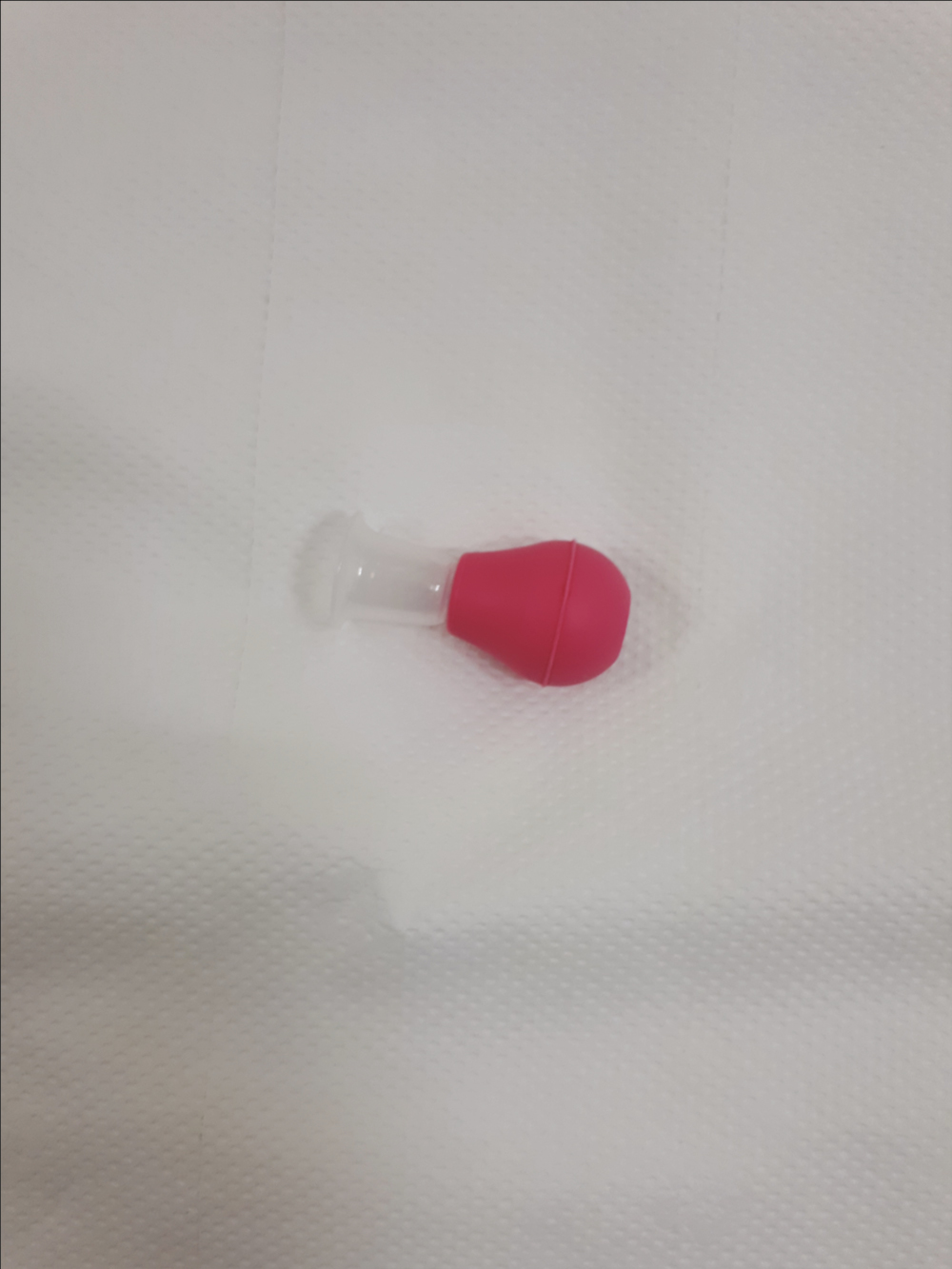
- Suction device
We recommend the suctioning to be done by [Grades 2 and 3] patients for a minimum period of 6 months, although it is advisable to use it till the patients start breastfeeding. [Grade 1] inverted nipples need not do suctioning.
RESULTS
We had 11 cases of nipple inversion, of which 5 cases were bilateral. None of our cases had a recurrence. Minimum follow-up period was 1 year. One case had external stitch infection which healed with antibiotics.
Typical repair of unilateral inverted nipple is shown in Figure 2 (preoperative) and Figure 3 (postoperative).
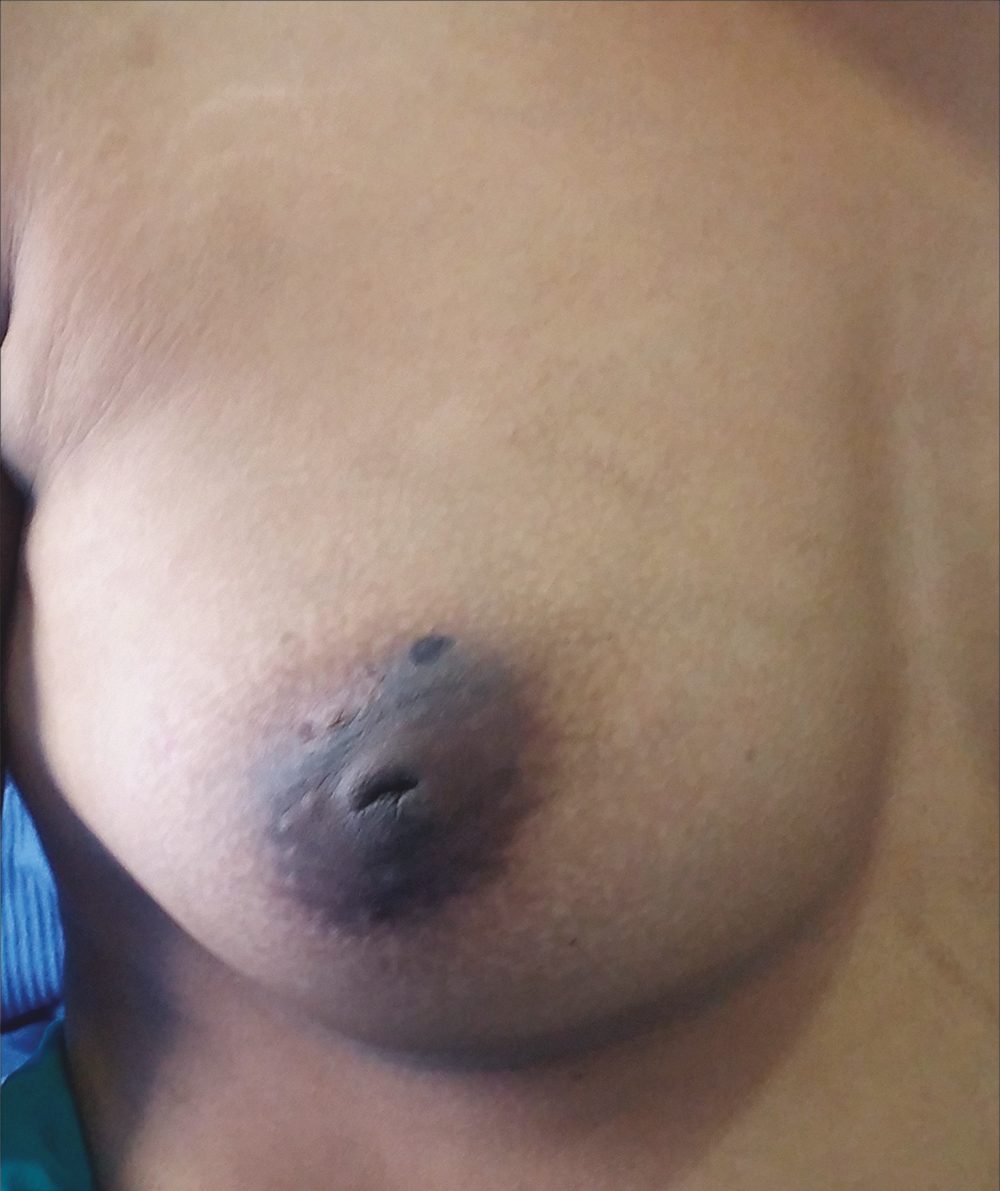
- Preoperative photo of unilateral inverted nipple
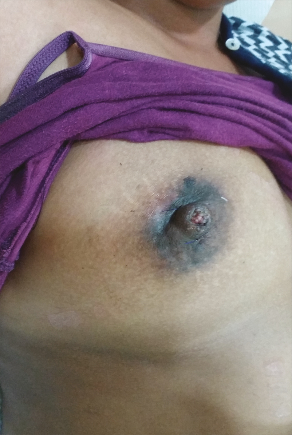
- Postoperative photo of unilateral inverted nipple
Typical repair of bilateral inverted nipple is shown in Figure 4 (preoperative) and Figure 5 (postoperative). Table 1 shows our demographic data.
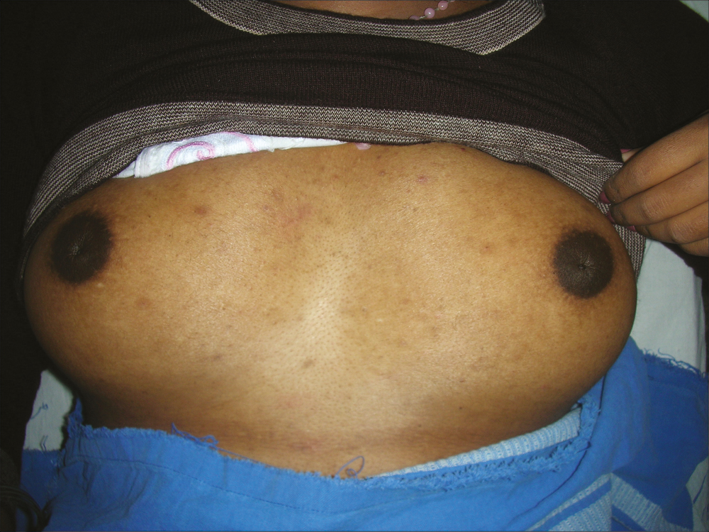
- Preoperative photo of bilateral inverted nipple
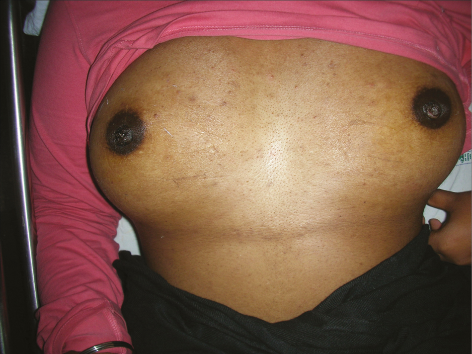
- Postoperative photo of bilateral inverted nipple
| Age | U/L OR B/L | Complication | Grade |
|---|---|---|---|
| 20 | u/l | Nil | 2 |
| 23 | b/l | Nil | 3 |
| 19 | u/l | Stitch infection | 2 |
| 18 | u/l | Nil | 2 |
| 26 | b/l | Nil | 1 |
| 23 | u/l | Nil | 3 |
| 20 | b/l | Nil | 3 |
| 25 | b/l | Nil | 3 |
| 22 | u/l | Nil | 2 |
| 26 | u/l | Nil | 2 |
| 24 | b/l | Nil | 3 |
DISCUSSION
Several methods have been described for the correction of congenital inverted nipple.
However, most of these methods involve dermal flaps or turnover flaps which can affect the lactation as injury to lactiferous ducts can occur.[6789101112]
Also most of these methods have no mention of etiology of inverted nipple. Also no particular mention of gravidity or parity of patient is mentioned.
This is of particular significance as feeding of the child can be affected in some of the methods as lactiferous ducts can get damaged during correction of inverted nipple.
Our method aimed at treatment of congenital inverted nipple in nulliparous women where minimal manipulation of lactiferous ducts is done. Our method has been particularly effective in preventing recurrence. We attribute it to use of nonabsorbable material for pursestring stitch and also strict adherence to postoperative suctioning using the suction device. This technique differs from article by Gould et al.,[5] where absorble material has been used as suture material and had a recurrence rate of 7%. Another difference is that in our postoperative protocol we recommend the use of suction device for [Grades 2 and 3] inverted nipples for 6 months to prevent recurrence.
CONCLUSION
We recommend this simple technique of pursestring technique for correction of congenital inverted nipple. The advantage of this technique is that it can be done as an outpatient procedure under local anesthesia. It has no effect on future lactation of the patient and has minimal chances of recurrence with strict adherence to postoperative suctioning protocol.
Statements of human or animal rights or ethical approval
This study does not involve any experimental study on humans and is a therapeutic study.
Informed consent
Informed consent has been taken from all patients.
Financial support and sponsorship
Nil.
Conflicts of interest
There are no conflicts of interest.
Declaration of patient consent
The authors certify that they have obtained all appropriate patient consent forms. In the form the patient(s) has/have given his/her/their consent for his/her/their images and other clinical information to be reported in the journal. The patients understand that their names and initials will not be published and due efforts will be made to conceal their identity, but anonymity cannot be guaranteed.
All videos available online www.jcasonline.com
REFERENCES
- Inversion of the human female nipple, with a simple method of treatment. Plast Reconstr Surg. 1974;54:564-9.
- [Google Scholar]
- The nipple: A simple intersection of mammary gland and integument, but focal point of organ function. J Mammary Gland Biol Neoplasia. 2013;18:121-3.
- [Google Scholar]
- inverted nipple repair revisited: A 7-year experience. Aesthet Surg J. 2015;35:156-64.
- [Google Scholar]
- Correction of inverted nipples by strong suspension with areola based dermal flaps. Plast Reconstr Surg. 2009;123:1131-2.
- [Google Scholar]
- Simple technique for correction of inverted nipple. Plast Reconstr Surg. 1980;65:504-6.
- [Google Scholar]
- Correction of inverted nipple using strut reinforcement with deepithelialized triangular flaps. Plast Reconstr Surg. 1998;102:1253-8.
- [Google Scholar]
- Correction of inverted nipple: An alternative method using two triangular areolar dermal flaps. Ann Plast Surg. 2003;51:636-40.
- [Google Scholar]
- Correction of inverted nipples by strong suspension with areola-based dermal flaps. Plast Reconstr Surg. 2007;120:1483-6.
- [Google Scholar]
- Surgical correction of inverted nipples using the modified Namba or Teimourian technique. Plast Reconstr Surg. 2004;113:328-36; discussion 337-328.
- [Google Scholar]
- The application of de-epithelialised “turn-over” flaps to the treatment of inverted nipples. Br J Plast Surg. 1984;37:253-5.
- [Google Scholar]


