Translate this page into:
Quantitative trichoscopic analysis of hair density in people attending tertiary care hospital – A cross-sectional study
*Corresponding author: Shraddha Uday Katruwar, Department of Dermatology, Ashwini Rural Medical College, Hospital and Research Centre, Solapur, Maharashtra, India. shraddhakatruwar@gmail.com
-
Received: ,
Accepted: ,
How to cite this article: Katruwar SU, Shah S. Quantitative trichoscopic analysis of hair density in people attending tertiary care hospital – A cross-sectional study. J Cutan Aesthet Surg. 2024;17:194-7. doi: 10.25259/jcas_66_23
Abstract
Objectives:
Hair is important to trace evidence commonly encountered in almost all criminal cases. Forensic anthropologists routinely compare the morphological characteristics of the hair samples to determine a transfer. Assessment of parameters such as hair density, hair diameter, follicular units, and empty hair follicle/yellow dots are useful for the diagnosis of various hair disorders, monitoring treatment outcomes, and research purposes. The study aimed to analyze the relationship between age and hair density.
Material and Methods:
The present cross-sectional observational study was carried out at the Department of Dermatology at a tertiary care center. All the subjects attending the Dermatology Outpatient Department fulfilling inclusion criteria were studied. Data were collected after the approval from the Institutional Ethical Committee. The procedure was explained and a written consent was taken. Each patient’s hair was parted in the middle and images from a point of roughly 5 inches from the patient’s glabella were taken. The parameters such as hair thickness, hair density, hair count, follicular units, and empty hair follicles/yellow dots were noted.
Results:
In the present study, the majority of the study subjects belonged to the age group 26–35 years (38%), followed by 18–25 years (34.7%), 36–45 years (20.7%), and least in the age group of 46–55 years (6.7%). In the present study, the mean unit density in males was 104.78 ± 14.33 and that of females was 108.36 ± 17.60.
Conclusion:
Mean hair density varies concerning age group in the study population.
Keywords
Hair
Age
Trichoscopic
Scalp
INTRODUCTION
Mechanical properties of the hair are attributed to the cortex, which forms the bulk of the fiber. Hair is important to trace evidence commonly encountered in almost all criminal cases. Forensic anthropologists routinely compare the morphological characteristics of the hair samples to determine a transfer.1 However, many questions such as how populations can be analyzed and possibly distinguished based on the morphology and appearance of their hair remain unanswered. Assessment of parameters such as hair density, hair diameter, follicular units, and empty hair follicle/yellow dots is useful for the diagnosis of various hair disorders, monitoring treatment outcomes, and research purposes.2 The hair parameters can be assessed using a trichoscopy. Trichoscopy is a videodermoscopy of hair and scalp.3 It is a new method that has been developed for hair image analysis which is a non-invasive, easy-to-use, and less time-consuming method, and provides accurate results. It provides ×20–70 magnification of the images.4
However, there is a dearth of data on the Asian population using the same evaluation procedure. The purpose of this study was to analyze hair density concerning age groups, as well as to identify the effect of sex and aging on these values.
MATERIAL AND METHODS
The present cross-sectional observational study was carried out under the Department of Dermatology at a tertiary care center. All the subjects attending the Dermatology Outpatient Department (OPD) of Tertiary Rural Health Care Center fulfilling inclusion criteria were studied. Data were collected after the approval from the Institutional Ethical Committee. The procedure was explained and a written consent was taken and signed by each patient participating in the study in the language best understood by the patient.
Inclusion criteria
All patients in the age group of 18–60 years attending dermatology OPD in a tertiary care hospital were included in the study.
Exclusion criteria
The following criteria were excluded from the study:
Self-reported hair loss within 6 months before the study.
Self-reported uncontrolled systemic conditions such as diabetes mellitus, thyroid disorders, blood pressure, connective tissue disorders, and cancer.
Patients undergoing treatment for scalp disorders such as microsporidiosis, various types of alopecias, and hair shaft abnormalities.
Positive hair pull test.
Autoimmune diseases like alopecia areata.
Nutritional deficiencies such as iron deficiency anemia and megaloblastic anemia.
Patients with abnormal scalp findings on physical examination. Any scalp abnormalities detected on trichoscopy.
Sample size was calculated by the formula = Z 2 × S2/(M × D)2
Where Z = value of confidence level at 95% = 1.96
S = Standard deviation from previous study = 12.80 (frontal area of scalp)2
M = Mean from previous study = 154.30 (frontal area of scalp)2
D = Absolute precision = 1%. Thus, a total of 300 cases were studied using simple random sampling.
Data collection procedure
Each patient’s hair was parted in the middle and images from a point of roughly 5 inches from the patient’s glabella were taken. Thereafter, the images were taken 3 inches to the right, left, and backward thereby covering the whole scalp. The images were taken at 50-fold magnification and the area covered is 0.238 sq. cm. The parameters such as hair thickness, hair density, hair count, follicular units, and empty hair follicles/yellow dots were noted in the pro forma.
Statistical analysis
Data is entered into a Microsoft Excel sheet and analyzed using the Statistical Package for the Social Sciences 24.0 version IBM USA. Qualitative data were expressed in terms of proportions. Quantitative data were expressed in terms of mean and standard deviation. Association between two qualitative variables was seen using Chi-square/Fisher’s exact test. P < 0.05 was considered as significant.
RESULTS
In the present study, the majority of the study subjects belonged to the age group 26–35 years (38%), followed by 18–25 years (34.7%), 36–45 years (20.7%), and least in the age group of 46–55 years (6.7%). The average age is 30.4 ± 8.4 with a minimum of 18 years and a maximum of 54 years [Table 1].
| Age in years | Frequency | Percentage |
|---|---|---|
| 18-25 | 104 | 34.7 |
| 26-35 | 114 | 38 |
| 36-45 | 62 | 20.7 |
| 46-55 | 20 | 6.7 |
| Total | 300 | 100 |
It was observed that the majority of study subjects were female (53.3%) and male (46.7%).
The mean hair count terminal on males was 40.03 ± 5.22 and that of females was 40.12 ± 4.66. There is no significant difference in hair count terminal between males and females (P = 0.89). In turn, the hair count terminal is almost the same in both males and females [Table 2].
| Gender | Frequency | Percentage |
|---|---|---|
| Female | 160 | 53.3 |
| Male | 140 | 46.7 |
| Total | 300 | 100 |
There is a significant negative correlation between age with hair density terminal (r = −0.144, P = 0.01), whereas there is a significant positive correlation between age with hair density vellus (r = 0.183, P = 0.001) [Table 3].
| r-value | P-value | |
|---|---|---|
| Hair density terminal (/cm2) | -0.144 | 0.01 |
| Hair density vellus (/cm2) | 0.183 | 0.001 |
| Mean thickness μm) | -0.094 | 0.10 |
| Units density | -0.027 | 0.64 |
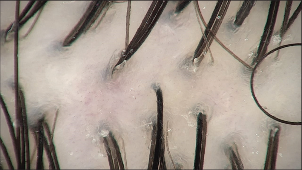
Trichoscopic image shows the follicular unit with 2–3 hairs, terminal hair, vellus hair, honeycomb pigment network, and white dots [Figure 1].

- Hair follicle.
DISCUSSION
In the present study, the majority of the study subjects belonged to the age group 26–35 years (38%), followed by 18–25 years (34.7%), 36–45 years (20.7%), and least in the age group of 46–55 years (6.7%). Leerunyakul and Suchonwanit,5 in 2020, conducted a study whose hair examination findings were normally evaluated. Two hundred and thirty-nine subjects participated in this study, of whom 79 were male and 160 were female with an average age of 37.9 years. Our findings are not consistent with the above study since the mean age was higher and may be due to ethnic differences.
In the present study, the mean ± standard deviation (SD) of hair parameters were as follows: hair density terminal was 168.62 ± 20.80, hair density vellus 26.08 ± 10.65 and mean thickness 0.06 ± 0.007. Hu et al.6 reported that the hair density, mean hair per follicle unit, and vellus hair ratio from 4-mm punch biopsy were 214.97 ± 48.73/cm2, 2.24 ± 0.30, and 10.48 ± 6.43%, higher than those from quantitative trichoscopy analysis (163.07 ± 28.17/cm2, 1.87 ± 0.25 and 6.60 ± 3.95%). The hair shaft diameter and terminal hair shaft diameter of the biopsy were 68.65 ± 8.00 µm and 73.77 ± 6.74 µm, less than those of trichoscopy (74.52 ± 8.02 µm and 77.87 ± 7.53 µm). The differences were statistically significant (P < 0.05). Our findings are not consistent with the above study which may be due to ethnic differences. Leerunyakul and Suchonwanit5 reported comparisons of hair diameter as follows: Frontal 81.3 ± 4.4 and 81.2 ± 5.1, vertex-81.1 ± 6.5 and 80.7 ± 6.3, temporoparietal −80.2 ± 5.8 and 80.3 ± 4.7, and occipital −80.1 ± 3.6 and 80.1 ± 6.9.
In the present study, the mean unit density in males was 104.78 ± 14.33 and that of females was 108.36 ± 17.60. Chen et al.7 reported that the average occipital hair shaft diameter and single hair follicle unit ratio of 35 normal Chinese females from quantitative trichoscopic analysis were 68.34 ± 7.70 µm and 13.47 ± 8.74%, which were slightly lower than this study (74.52 ± 8.02 µm and 33.77 ± 12.78%). However, the occipital vellus hair ratio reported by Chen was 8.88 ± 4.23%, higher than 6.60 ± 3.95% in this study. The distinction may be caused by different magnification and count modes.
The previous research in Asians utilizing computer-assisted phototrichogram found a reduction in hair density in normal Japanese females beyond the age of 40.8 In Korean studies, a similar observation was made.9 When hair diameter was compared between age groups, Japanese girls in their 20s had a different hair diameter than those in their 40s. However, while Kim et al. observed a decrease in hair diameter beginning in the 40s, this was not statistically significant.9 Our study found that hair density declined with age, but the reduction was not great enough to be statistically significant until participants were in their 60s. Our findings also revealed that aging causes hair follicular stem cells to senescence and lose their function.
It is common knowledge that the frequency of hair loss rises with age. Several earlier research demonstrated that age variations affect hair density in the absence of hair loss.
Because our study was conducted at a single tertiary care center, it may not be representative of the overall community. To overcome this problem, another nationwide cross-sectional study with stratification could be conducted. As a result, a large-scale study with a diverse sample size should be conducted in the future to assess the pattern of hair density and different age groups among different ethnicities.
CONCLUSION
In the present study, mean hair density varies concerning age group in the study population. A deeper awareness of hair density differences on the scalp can also be relevant for evaluating and selecting hair transplantation treatments for individuals. Furthermore, these disparities are significant because being aware of ethnic differences in hair density can affect the capacity to effectively diagnose hair diseases, evaluate response to therapy, and undertake hair-related research in these patients.
Ethical approval
The research/study approved by the Institutional Review Board at Ashwini Rural Medical College, Hospital and Research Centre, Kumbhari, Solapur, Approval No.-ARMCH/IEC/26/2021, Date - 07-01-2021.
Declaration of patients consent
The authors certify that they have obtained all appropriate patient consent.
Conflicts of interest
There are no conflicts of interest.
Use of artificial intelligence (AI)-assisted technology for manuscript preparation
The authors confirm that there was no use of artificial intelligence (AI)-assisted technology for assisting in the writing or editing of the manuscript and no images were manipulated using AI.
Financial support and sponsorship
Nil.
References
- Chemical and physical behavior of human hair (5th ed). Berlin, Heidelberg: Springer Berlin Heidelberg; 2012.
- [Google Scholar]
- A study of variations in some morphological features of human hair. J Punjab Acad Forensic Med Toxicol. 2002;2:1-6.
- [Google Scholar]
- Malaysia's three major ethnic group preferences in creating a Malaysian garden identity. Aust Geogr. 2013;44:197-213.
- [CrossRef] [Google Scholar]
- A study on scalp hair health and hair care practices among Malaysian medical students. Int J Trichology. 2017;9:58-62.
- [CrossRef] [PubMed] [Google Scholar]
- Evaluation of hair density and hair diameter in the adult Thai population using quantitative trichoscopic analysis. Biomed Res Int. 2020;2020:2476890.
- [CrossRef] [PubMed] [Google Scholar]
- Comparison between trichoscopic and histopathological evaluations of hair parameters. Clin Cosmet Investig Dermatol. 2022;15:843-9.
- [CrossRef] [PubMed] [Google Scholar]
- A two-step mechanism for stem cell activation during hair regeneration. Cell Stem Cell. 2009;4:155-69.
- [CrossRef] [PubMed] [Google Scholar]
- Characteristic features of Japanese women's hair with aging and with progressing hair loss. J Dermatol Sci. 2007;45:93-103.
- [CrossRef] [PubMed] [Google Scholar]
- Characteristic features of ageing in Korean women's hair and scalp. Br J Dermatol. 2013;168:1215-23.
- [CrossRef] [PubMed] [Google Scholar]


