


Translate this page into:
Solitary Neurofibroma over Lower Lip: A Rare Manifestation
Address for correspondence: Dr. B. Spoorthy, Department of Dermatology, Venereology and Leprosy, Vydehi Institute of Medical Sciences and Research Centre, 82, Near BMTC 18th Depot, Vijayanagar, Nallurhalli, Whitefield, Bengaluru 560066, Karnataka, India. E-mail: bspoorthy94@gmail.com
This is an open access journal, and articles are distributed under the terms of the Creative Commons Attribution-NonCommercial-ShareAlike 4.0 License, which allows others to remix, tweak, and build upon the work non-commercially, as long as appropriate credit is given and the new creations are licensed under the identical terms.
This article was originally published by Wolters Kluwer - Medknow and was migrated to Scientific Scholar after the change of Publisher.
Abstract
Abstract
Neurofibromas are common nerve sheath tumors, occurring either sporadically or associated with Von Recklinghausen’s disease. Only 6.5% of solitary lesions are seen to involve the oral cavity without any features of neurofibromatosis type 1 (NF-1). It presents as a soft, skin-colored nodule with a characteristic buttonhole invagination. Histologically, it is an unencapsulated lesion consisting of proliferated neural elements, with a background of mucin and mast cells. Surgical excision is the treatment of choice. A 49-year-old female patient presented with a 35-year history of single, asymptomatic lesion over the lower lip, with no features of NF-1. On the basis of the history, histopathological findings, and dermoscopy, a diagnosis of neurofibroma was made and the lesion was excised, with no recurrence over a period of 1 year. The present case report has been reported for its interesting presentation and unusual site of involvement.
Keywords
Lower-lip
neurofibroma
solitary

INTRODUCTION
Neurofibromas are the most common benign peripheral nerve sheath tumors, composed of Schwann cells, perineural-like cells, and fibroblasts.[1] They can occur either as localized lesions or a part of generalized syndrome of neurofibromatosis type 1 (NF-1). Solitary neurofibromas develop as a well-circumscribed mass along a peripheral nerve but are unencapsulated. The skin is the most commonly involved site, rarely occurring in the oral mucosa.[2] Most of the neurofibromas present in the second to third decade of life, with no sex predilection.
Only 25% of cases are seen to be involving the head and neck regions. Out of which 6.5% of patients present with oral lesions. Tongue is the most common site of involvement, with rare presentation over the lips.[3]
Microscopically, the neurofibromas of sporadic or syndromic origin are similar, composed of spindle cells with myxoid stroma and scattered mast cells. They are immunopositive for S-100 protein.
Here we report a case of solitary neurofibroma over the lower lip in the absence of any signs of NF-1.
CASE REPORT
A 49-year-old female patient presented with a 35-year history of solitary swelling over the lower lip nonprogressive in size or number. It was not associated with any pain, itching, or bleeding. No similar lesions were noted elsewhere on the body.
There was no spontaneous resolution seen over years. There was no significant medical or family history.
Physical examination revealed a solitary skin-colored, well-defined, firm, compressible, nontender nodule of 0.5 cm × 0.5 cm size present over the left side of lower lip, with no secondary changes. No café au lait spots, axillary freckling, skeletal abnormalities, and Lisch nodules were noted. The findings of systemic examination were unremarkable [Figure 1].
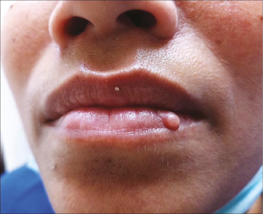
On dermoscopy, a yellowish-white mass with an overlying vascular network in a reticular pattern was seen [Figures 2 and 3].


A provisional diagnosis of mucocele, fibroma, or lymphatic hemangioma was made, and the lesion was excised completely.
On histopathological examination, an encapsulated lesion composed of bland spindle cells arranged in a storiform pattern was seen in the dermis. It was interspersed with collagen bundles and scattered mast cells, with hyalinized thickened blood vessels [Figures 4 and 5].


On the basis of clinicopathological correlation, a diagnosis of neurofibroma (a spindle cell neoplasm) was made.
DISCUSSION
Neurofibromas are benign tumors of the nerve sheath, which can occur sporadically or as a component of neurofibromatosis. When one or two such lesions are found, they are usually sporadic in nature without any systemic manifestations. However, more than three neurofibromas point toward neurofibromatosis, with other cutaneous, skeletal, and neural manifestations.[4]
Neurofibromas in the head and neck region can be either cutaneous, mucosal, or deep neural bundles, usually related to facial, glossopharyngeal, or vagus nerves.
Oral cavity neurofibromas usually occur as a part of syndrome, that is, NF-1 or NF-2. Solitary neurofibromas are rare in the oral cavity with a prevalence of 4%–7%. On the basis of the literature, tongue is the most common site followed by palate, mandibular ridge, maxillary ridge, buccal mucosa, and gingiva (in the order of frequency). They appear as pedunculated or sessile nodules, characterized by slow growth and asymptomatic nature.[45]
Solitary cutaneous neurofibromas are not associated with axillary freckling, café au lait spots, Lisch nodules, scoliosis, or any family history of NF-1.
Differential diagnosis includes mucocele, fibroma, dermatofibroma, lipoma, and lymphatic hemangioma. Mucoceles are cysts due to mucus accumulation, presenting as bluish, soft, and translucent swellings, often preceded by trauma. They usually resolve spontaneously. Lipomas are yellowish, soft swellings composed of fat tissue, which show the characteristic slip sign. Fibromas are nodules with hard consistency, sessile, and located often on the buccal mucosa.
Microscopically, they are unencapsulated spindle cell tumors with a bland appearance, restricted to corium or extending till subcutis. They are arranged in various patterns such as thin fascicles, storiform, or in haphazard fashion. Surrounding dermis shows myxoid changes with few scattered mast cells. This appearance has been called “shredded carrot” appearance. In 85%–100% of cases, immunostaining with S-100 confirms its neural origin.[67]
On dermoscopy, yellow-to-skin-colored background is the most commonly observed global pattern. However, focal dermoscopy shows exaggerated skin markings and pigment-like pattern frequently. Dermatofibroma shows comedo-like openings with irregular crypts.[7] Mucocele shows either reticular or hairpin-like vessels on an erythematous base.[8]
Solitary oral neurofibromas are stable in nature, which seldom shows a malignant transformation into a sarcoma. Treatment modalities include physical removal like surgery, radiofrequency ablation, CO2 laser, and electrodesiccation. If admixed with nerves, preservation of nerve is of utmost importance.[910] Recently, FDA has approved Selumetinib (MEK inhibitor) for symptomatic neurofibromas.
CONCLUSION
Solitary neurofibromas over the lips are an unusual presentation and are one of the differential diagnoses of solitary nodules. They usually do not show any other features of NF-1 or NF-2. They are characterized by slow growth and asymptomatic nature. Excision is the treatment of choice, with minimal recurrence rates.
Providing insight into this cutaneous disorder is important for patient education and positive outcomes to affected individuals.
Financial support and sponsorship
Nil.
Conflicts of interest
There are no conflicts of interest.
Declaration of patient consent
The authors certify that they have obtained all appropriate patient consent forms. In the form the patient(s) has/have given his/her/their consent for his/her/their images and other clinical information to be reported in the journal. The patients understand that their names and initials will not be published and due efforts will be made to conceal their identity, but anonymity cannot be guaranteed.
REFERENCES
- Solitary neurofibroma of the floor of the mouth: Rare localization at lingual nerve with intraoral excision. BMC Oral Health. 2019;19:197.
- [Google Scholar]
- A rare solitary neurofibroma of the philtrum of upper lip. Dent Oral Maxillofac Res. 2020;6:1-3.
- [Google Scholar]
- Sporadic and multiple neurofibromas in the head and neck region: A retrospective study of 33 years. Clin Oral Investig. 2007;11:165-9.
- [Google Scholar]
- Solitary neurofibroma at the base of the tongue: A rare presentation. Indian J Dermatol. 2015;60:497-9.
- [Google Scholar]
- Solitary neurofibroma of the floor of the mouth: A case report. Case Rep Otolaryngol. 2011;2011:967896.
- [Google Scholar]
- Cutaneous tumors and pseudotumors of the head and neck. In: Gnepp D, Bishop J, eds. Gnepp’s diagnostic surgical pathology of the head and neck (3rd ed). Elsevier; 2020. p. :1012-97.
- [Google Scholar]
- Dermoscopic features of solitary neurofibroma: A retrospective analysis of 32 cases. Australas J Dermatol. 2020;61:e406-9.
- [Google Scholar]
- Dermoscopy of oral mucocele: Three types of extravasation mucoceles. Turk J Med Sci. 2020;50:96-102.
- [Google Scholar]
- Nonsyndromic solitary neurofibromas in the oral cavity: Case series and literature review. J Indian Acad Oral Med Radiol. 2016;28:52-6.
- [Google Scholar]
- Management of cutaneous neurofibroma: Current therapy and future directions. Neurooncol Adv. 2020;2:i107-16.
- [Google Scholar]