Translate this page into:
Surgical Excision and Primary Closure of Venous Lake of Upper Eye Lid for Better Cosmetic Outcome: A Case Report
Address for correspondence: Dr. Yogesh M. Bhingradia, Department of Dermatology, Shivani Skin Care and Cosmetic Clinic, Sarthi Doctor House, Fourth Floor, Hira Bag, Varachha Road, Surat 395006, Gujarat, India. E-mail: yogeshbhingradia@gmail.com
This article was originally published by Wolters Kluwer - Medknow and was migrated to Scientific Scholar after the change of Publisher.
Abstract
Abstract
Venous lakes (VLs) are fairly common vascular lesions caused by dilatation of the localized vessels on sun-damaged skin. They are usually asymptomatic; however treatment is opted to improve psychological distress caused by cosmetic disfigurement and occasionally to prevent bleeding. Multiple treatment modalities such as cryosurgery, carbon dioxide laser, pulse dye laser, sclerotherapy, and electrocoagulation have been mentioned in literatures with varying degrees of success and specific complications. Hereby, we present a case of a 40-year-old female with VL on upper eyelid treated successfully with surgical excision with better cosmesis.
Keywords
Better cosmetic outcome
surgical excision
venous lake
INTRODUCTION
Venous lakes (VLs) are fairly common vascular lesions caused by dilatation of the localized vessels, with male: female ratio being 1.5:1. They are sharply demarcated, round to oval, dark purple to blue papule, mostly present on lips, followed by face and ears in elderly population. Usually, they are single in number but occasionally multiple lesions can occur.[1] Histologically, they are lined by a single layer of flattened endothelial cells and a thick wall of fibrous tissue is located in the upper dermis. The lesion is thought to occur as a result of the deterioration in connective tissue in vascular adventitia and in the dermis.[23] VLs are benign and usually asymptomatic but occasionally can bleed on minor trauma, which can be cosmetically disturbing.
Multiple treatment modalities such as cryosurgery, carbon dioxide laser, pulse dye laser (PDL), long pulse Nd:YAG laser, sclerotherapy, and electrocoagulation have been mentioned in literatures.[456] Various laser therapies have been tried with varying results and specific complications, giving poor cosmesis. The use of PDL for treatment of VLs has comparatively lower risk of scarring than other types of lasers. But results with PDL treatment for VLs are variable and also demanded multiple sessions. The long-pulsed Nd:YAG laser has also been reported effective for vascular lesions on face and neck with variable results and long-term follow up. So, keeping in mind the patient compliance and better cosmetic outcome, we opted for surgical excision of the VL.
CASE REPORT
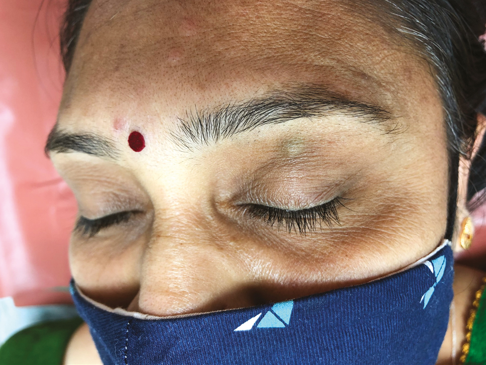
A 40-year-old female patient visited our clinic for removal of the bluish purple color lesion on upper eye lid since several years. The lesion was asymptomatic, soft, compressible papule of around 8 mm in diameter situated 1 cm below the middle third of the left eyebrow [Figure 1]. No similar lesions were noted elsewhere on the body. Dermoscopy was done, which showed globules/clods with purple/blue coloration, which is an indicator for VL.

- VL on left upper eyelid
MATERIALS AND METHODS
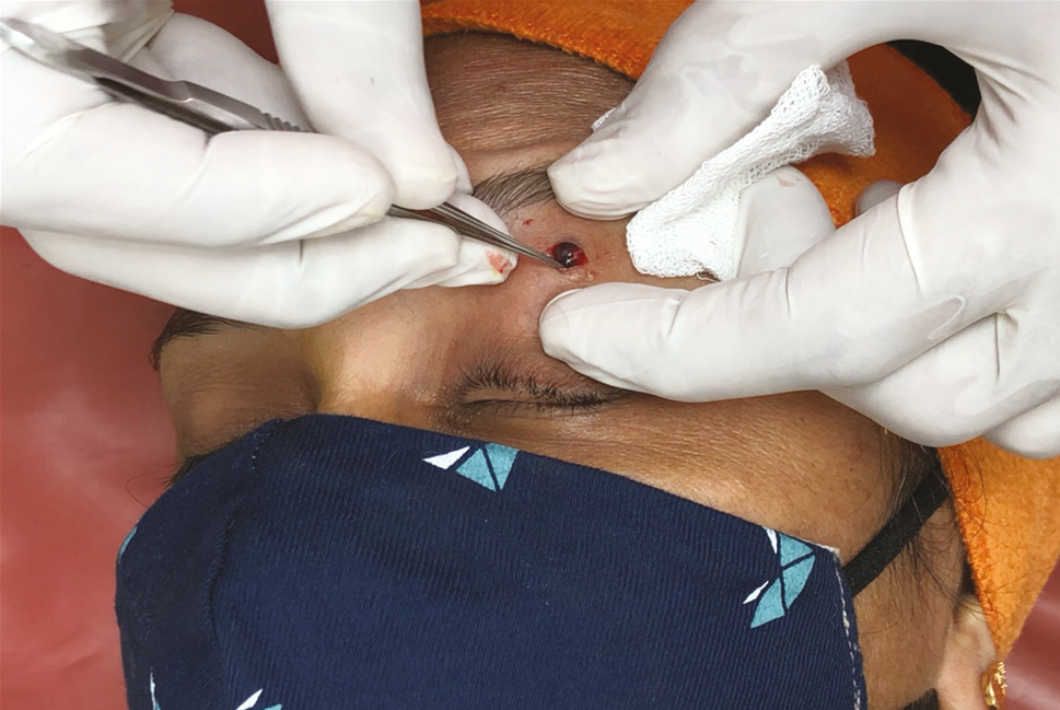
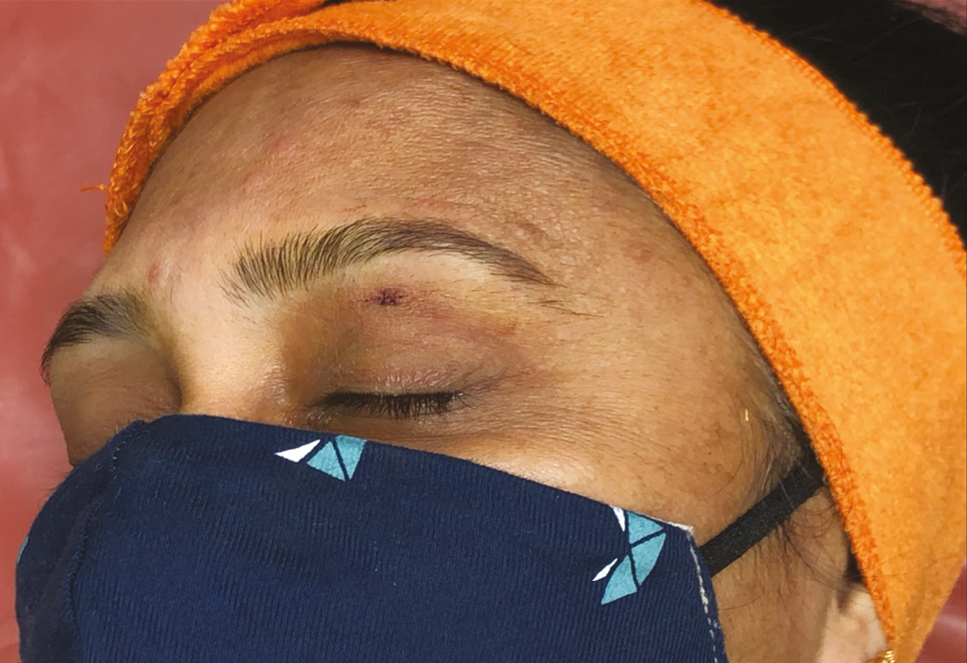
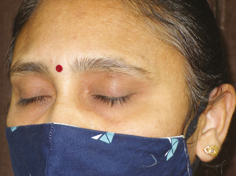
After obtaining the informed written consent and preprocedural photographs for record, the patient was prepared for surgical excision keeping all aseptic precautions in mind. The site was draped with a sterile sheet and thoroughly cleaned it with spirit, betadine, and normal saline. Local anesthesia was given using insulin syringe, and an incision was made with a 15 no. blade using the single stroke method. VL was removed by holding it using toothed forceps with blunt dissection, and bleeding points were cauterized by RF cautery [Figure 2]. The primary closure was done using non-absorbable 6-0 simple interrupted sutures [Figure 3]. After applying topical antibiotics, the site was packed with sterile gauze and skin tape. Excised tissue was sent for histopathological examination, which showed the dilated lumen lined by a single layer of flattened endothelial cells and the lumen filled with erythrocytes, which confirmed the diagnosis of VL. Sutures were removed after 7 days, and the follow-up visit after a month showed good cosmetic scar [Figure 4].

- Removal of VL using a toothed pair of forceps

- Closure using non-absorbable 6-0 Ethilon, simple interrupted sutures

- After 1 month of suture removal
DISCUSSION
VLs are usually common on face due to constant sun exposure, especially around lips. It has been previously treated by multiple laser therapy, infrared coagulation, and cryotherapy. Chemotherapy has also been previously tried and reported. Although a patient with a VL is usually free of symptoms, it must sometimes be treated because of recurrent traumatic bleeding, as well as due to the cosmetic concerns.
Laser therapy may result in skin atrophy, hyperpigmentation, a depressed scar, and increased surgical cost, thus resulting in poor patient compliance.[27,8] PDL and long-pulsed Nd:YAG laser are being used for years to treat a variety of vascular lesions. The PDL induces selective destruction of cutaneous small vessels, and so it is considered the most effective device for the treatment of smaller and more superficial cutaneous vessels. The limitation of PDL is because of shallow depth of the penetration. For treatment of VLs, PDLs have resulted in the minimal scarring and adverse effects, but repetitive sessions are needed to achieve the complete resolution of the lesion. Long-pulsed 1064 nm Nd:YAG laser, however, provides greater depth of the penetration but low specificity. It penetrates deeply into the skin and also has greater affinity for much larger and deeper blood vessels. Owing to the deep penetration, long-pulsed Nd:YAG lasers have shown effectiveness in treating VLs, but with depressed scar formation.
Cryotherapy is also useful in the treatment of VLs, but multiple sessions are required and may also result in scarring and hyper–hypopigmentation.[9] Sclerosants used in sclerotherapy may produce allergic reactions, pain, burning, itching, ulceration, and swelling.
So, keeping in mind all the pros and cons of the various treatment options, we decided to go for surgical excision of the lesion. Surgical excision is a single session technique with least complications and better aesthetic outcome, giving better patient compliance.
Declaration of patient consent
The authors certify that they have obtained all appropriate patient consent forms. In the form the patient(s) has/have given his/her/their consent for his/her/their images and other clinical information to be reported in the journal. The patients understand that their names and initials will not be published and due efforts will be made to conceal their identity, but anonymity cannot be guaranteed.
Financial support and sponsorship
Nil.
Conflicts of interest
There are no conflicts of interest.
REFERENCES
- Dermatoses resulting from disorders of the veins and arteries. In: Rook’s textbook of dermatology Vol Vol. 3. (9th ed.). London: Wiley-Blackwell; 2016. p. :103-15.
- [Google Scholar]
- Evaluation of the treatment of venous lakes with the 595-nm pulsed-dye laser: A case series. Clin Exp Dermatol. 2007;32:148-50.
- [Google Scholar]
- Nd:YAG lasers (1,064 nm) in the treatment of venous malformations of the face and neck: Challenges and benefits. Lasers Med Sci. 2007;22:119-26.
- [Google Scholar]
- The effect of 0.5% sodium tetradecyl sulfate on a venous lake lesion. Ann Dermatol. 2008;20:179-83.
- [Google Scholar]
- Venous lakes of the lips successfully treated with a sclerosing agent 1% polidocanol: Analysis of 25 report cases. Int J Surg Case Rep. 2021;78:265-9.
- [Google Scholar]
- Venous lake of the lip treated with a sclerosing agent: Report of two cases. Dermatol Surg. 2003;29:425-8.
- [Google Scholar]
- Laser and light-based treatments of venous lakes: A literature review. Lasers Med Sci. 2016;31:1511-9.
- [Google Scholar]
- Venous lake of the lips treated using photocoagulation with high-intensity diode laser. Photomed Laser Surg. 2010;28:263-5.
- [Google Scholar]
- Venous lake treated by liquid nitrogen cryosurgery. Br J Dermatol. 1997;137:1018-9.
- [Google Scholar]


