Translate this page into:
Surgical Pearl: Simple Techniques for Achieving Hemostasis on Digital Lesions
Address for correspondence: Dr. Muhammed Mukhtar, Mukhtar Skin Centre, Katihar Medical College Road, Katihar 854105, Bihar, India. E-mail: drmmukhtar20@gmail.com
This article was originally published by Wolters Kluwer - Medknow and was migrated to Scientific Scholar after the change of Publisher.
Abstract
Abstract
The fingers are highly vascular sites which bleeds profusely during procedure. Penrose drain and gasket are not readily available in the clinic. For this problem, we advocated the use of hand gloves and cotton gauze to achieve haemostasis at the site of finger.
Keywords
Finger
finger gloves
gauze
knot 8
vascular lesions
PROBLEM
The fingers are highly vascular sites. On this, even small lesions bleed profusely during any procedure. For this problem, Penrose drain and gasket are commonly used to achieve hemostasis at the site.[12] However, these objects are not readily available in the clinic. We have used readily available medical objects in a different way to get hemostasis on the finger for a bloodless field during surgery.
SoLUTION
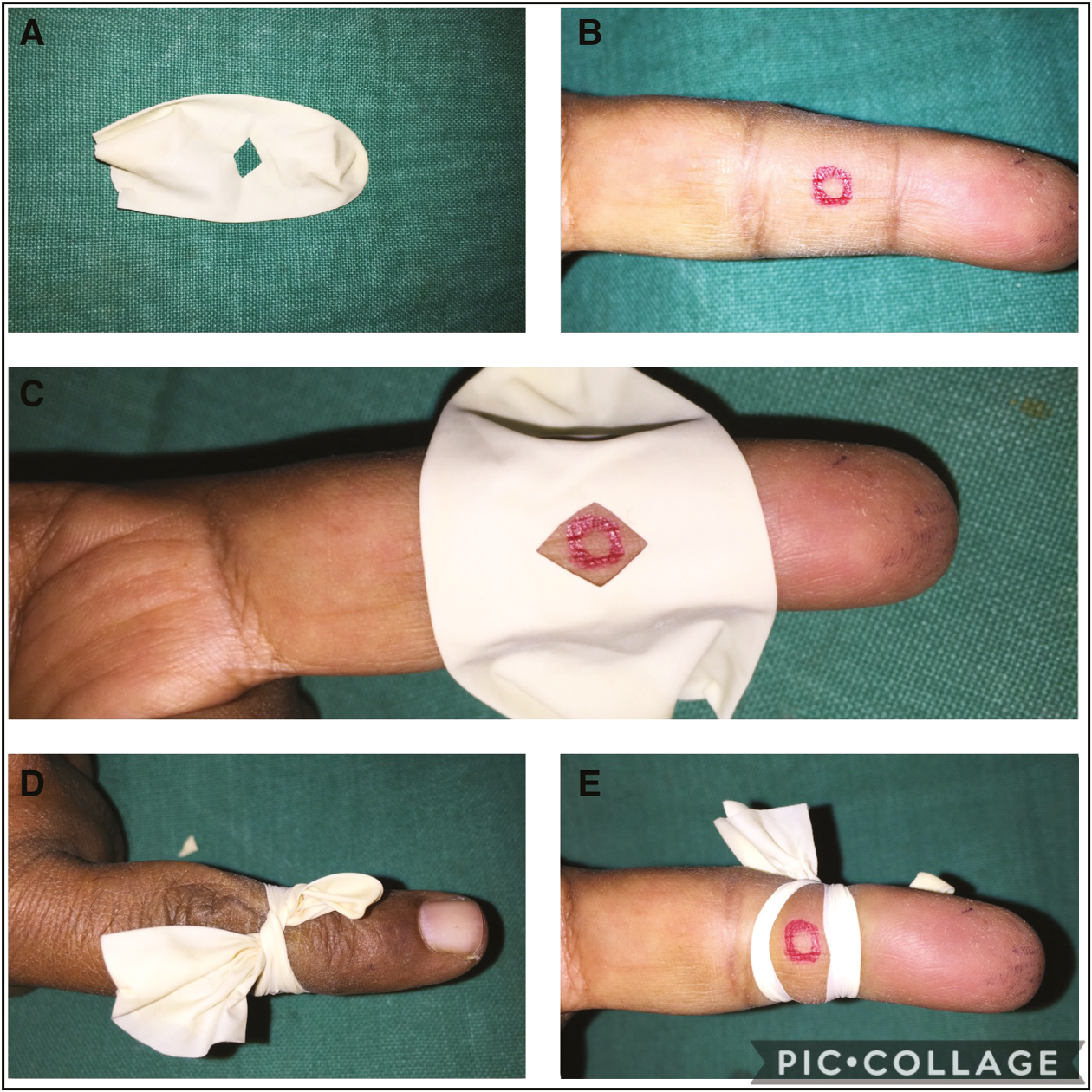
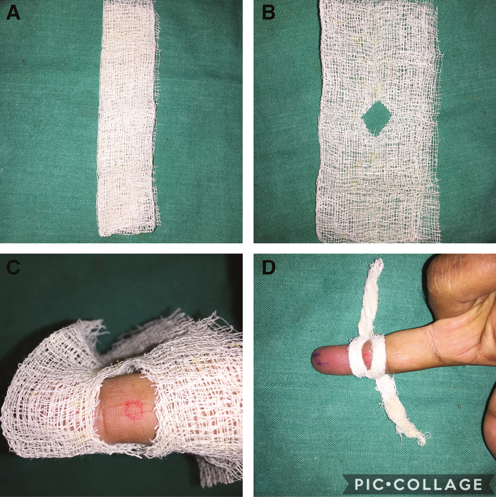
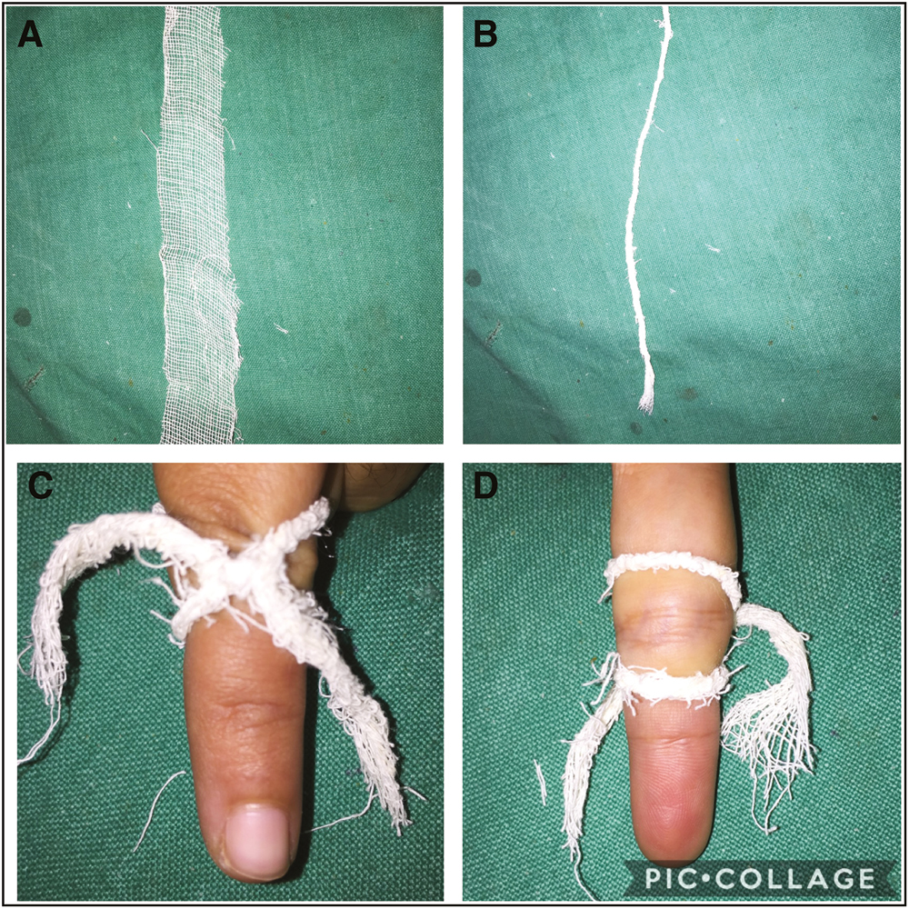
The blood circulation of fingers is supplied with a proper digital artery that runs along its both lateral volar borders. The pulse of digital artery can be felt at the border, and its blood pressure is about 80 mm Hg.[3] Hand gloves and cotton gauze have been used to achieve hemostasis at the operated site by “trapping” the digital lesions. For this, finger of a glove is cut, and a small hole (window) is made in the center. Subsequently, it is tied around the finger keeping the lesion within the hole as it is marked out [Figure 1A–E]. In place of finger gloves, a strip of gauze piece (6 × 2 inch, less expensive) can be used to make the site less vascular [Figure 2A–D]. The finger gloves and the gauze strip can be twisted out to make them like a small rope. This rope can be tied around the finger as a figure eight (8) knot for achieving hemostasis [Figure 3A–D]. After trapping the lesions for hemostasis, pulse of digital arteries becomes impalpable that can be confirmed by palpating the pulse and using a digital pulse oximeter, which can be made sure by pricking of the lesion. There are some limitations of these tapping methods. First, small lesions (granuloma pyogenicum, viral warts) can be trapped especially with finger gloves. Second, the trapping of the lesion or digit should be done for short time procedure (less than 20 min) to avoid acidosis and muscle fatigue in the finger. Third, the procedures requiring more than 20 min, then allow the finger to re-perfuse for 3–5 min.[4] Fourth, the trapping of digits should be avoided in patients with having pulse less or feeble pulse of digital artery.

- (A–E) A finger gloves with a hole (window) in the center is tied around the finger

- (A–D) A strip of gauze strip with a window is tied around finger

- (A–D) A rope of rolled strip of gauze is tied around finger as knot 8 to trap the lesion
Financial support and sponsorship
Nil.
Conflicts of interest
There are no conflicts of interest.
Declaration of patient consent
The authors certify that they have obtained all appropriate patient consent forms. In the form the patient(s) has/have given his/her/their consent for his/her/their images and other clinical information to be reported in the journal. The patients understand that their names and initials will not be published and due efforts will be made to conceal their identity, but anonymity cannot be guaranteed.
REFERENCES
- Trap technique for bloodless removal of digital pyogenic granuloma. J Am Acad Dermatol. 2020;83:e109-10.
- [Google Scholar]
- Noninvasive measurements of digital arterial pressure and compliance in man. Am J Physiol. 1977;233:H168-79.
- [Google Scholar]
- Effects of digital tourniquet Ischemia: A single centre study. Dermatol Surg. 2013;39:584-92.
- [Google Scholar]


