
Translate this page into:
Mondor’s Disease
Address for correspondence: Dr. Brett Neill, Division of Dermatology, University of Kansas Medical Center, 3901 Rainbow Blvd, Kansas City, Kansas 66160. E-mail: bcneill6@gmail.com
This is an open access journal, and articles are distributed under the terms of the Creative Commons Attribution-NonCommercial-ShareAlike 4.0 License, which allows others to remix, tweak, and build upon the work non-commercially, as long as appropriate credit is given and the new creations are licensed under the identical terms.
This article was originally published by Wolters Kluwer - Medknow and was migrated to Scientific Scholar after the change of Publisher.
Keywords
Dermatologic surgery
Mondor’s disease
superficial thrombophlebitis

A 44-year-old white female with no significant past medical history presented with a three-day history of mild tenderness of the left anterolateral thoracoabdominal wall. Twenty-three days earlier, she underwent an excision for a 6-mm nodular basal cell carcinoma of the left submammary area. Physical examination revealed three tender subcutaneous cord-like indurations without erythema that began below the well-healed surgical scar and continued toward the left lower quadrant of the abdomen [Figure 1]. Lifting the left arm above the head accentuated the appearance of the cords. Doppler ultrasonography revealed two noncompressible venous structures in the superficial subcutaneous tissues extending inferiorly over a length of a few centimeters, suggesting occlusive thrombi within small superficial veins just inferior to the surgical site. She was treated with nonsteroidal anti-inflammatory drugs and had a full recovery within seven weeks after presentation.
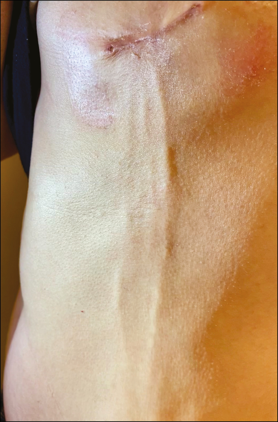
- Mondor’s disease. Three cord-like indurations on the anterolateral thoracoabdominal wall
DISCUSSION
Mondor’s disease (MD) was first described by Henri Mondor in 1939 as a superficial thrombophlebitis of a vein in the anterolateral thoracoabdominal wall. It is typically benign and self-limited, usually resolving in four to eight weeks without any specific treatment.[1] MD occurs most commonly in middle-aged women, with a female-to-male ratio ranging from 9:1–14:1.[1] No racial or ethnic propensity has been reported. The diagnosis is straightforward and based on physical examination. In cases where the clinical picture is not definitive, ultrasound is considered first-line treatment in the imaging evaluation of MD. The differential diagnosis includes thrombophlebitis migrans, infectious mastitis, Buerger disease, Behcet disease, and polyarteritis nodosa.[2] Forty-five percent of cases are idiopathic, 20% iatrogenic (including surgical chest operation, radiation, and hormone therapy), 22% traumatic (excessive physical activity and tight bra), and 5% related to breast cancer. Other risk factors include a hypercoagulable state and vascular disease. Nonsteroidal anti-inflammatory drugs can be used to relieve bothersome symptoms. Low-molecular-weight heparin or aspirin is not recommended in patients without underlying hypercoagulable state inciting superficial thrombophlebitis and associated MD.[2] The accurate identification of MD is important to first evaluate for the presence of underlying disease and to avoid performing any unnecessary invasive tests or treatment.
Declaration of patient consent
The authors certify that they have obtained all appropriate patient consent forms. In the form, the patient(s) has/have given his/her/their consent for his/her/their images and other clinical information to be reported in the journal. The patients understand that their names and initials will not be published and due efforts will be made to conceal their identity, but anonymity cannot be guaranteed.
Financial support and sponsorship
Nil.
Conflicts of interest
There are no conflicts of interest.


