Translate this page into:
Combined supraperiosteal microfat grafting and intradermal nanofat for the treatment of periorbital melanosis (dark circles)
*Corresponding author: Aniketh Venkataram, Department of Plastic Surgery, The Venkat Center for Skin ENT and Plastic Surgery, Bengaluru, Karnataka, India. anikethv@gmail.com
-
Received: ,
Accepted: ,
How to cite this article: Venkataram A. Combined supraperiosteal microfat grafting and intradermal nanofat for the treatment of periorbital melanosis (dark circles). J Cutan Aesthet Surg. 2025;18:114-9. doi: 10.25259/JCAS_10_2024
Abstract
Objectives:
Dark circles are one of the most common aesthetic concerns in India. While several treatment options exist, none address both volume deficiency and skin quality simultaneously. We felt that fat grafting and nanofat provided a novel treatment option to address both aspects of dark circles in one treatment.
Material and Methods:
Patient records were examined from 2017 to 2021. A total of 28 patients were identified as having undergone fat grafting and nanofat for dark circles specifically. The patients were analyzed for age, gender, volumes injected, and outcomes based on patient self-assessment.
Results:
A total of 36 patients underwent fat grafting ranging from the age of 20 to 40 (average 31). All procedures were done under local anesthesia as day care procedures. The volumes injected range from 2 cc/side to 8 cc/side, with an average of 4.36 cc. Using the Likert scale, 28 (77%) rated their results as very satisfied. Six (17%) rated it as satisfied. One rated it as neutral and two rated it as average, and underwent a second session of fat grafting where another 2 cc was injected per side.
Conclusion:
Fat grafting and nanofat are an exciting treatment option for the treatment of dark circles, which is usually regarded as a recalcitrant problem. It has the advantages of addressing both volume and skin quality, being a single-stage procedure, and producing comparatively long-lasting results.
Keywords
Dark circles
Fat grafting
Nanofat
Periorbital melanosis
Undereye rejuvenation
INTRODUCTION
Dark circles or periorbital melanosis is one the most common esthetic concerns in India. This term refers to the presence of a darker skin tone around the eyes, in particular, the undereye region. This condition is perceived to make an individual appear “more tired” or “more sleepy.”1
There are several proposed etiologies for dark circles including genetic, lifestyle, and inflammatory factors.2,3 The pathogenesis is linked to anatomical factors of reduced volume and physiological factors of increased pigmentation.
Treatment modalities thus far included options to replace volume such as fillers, and options to improve skin quality such as topicals and energy-based devices.2-4 While these techniques provide varying levels of success, none of them were able to address both aspects of dark circles, that is, volume and skin quality correction.
With this background, we felt that fat grafting and nanofat provided a novel treatment option to address both aspects of dark circles in one treatment. We present our experience with this technique.
MATERIAL AND METHODS
Patient records were examined from 2017 to 2021. A total of 36 patients were identified as having undergone fat grafting and intradermal nanofat for dark circles specifically. Patients who complained of age-related changes were excluded, as were patients for malar augmentation.
The patients were analyzed for age, gender, and volumes injected. Results were self-evaluated at 3 months post-procedure by the patients using the 5-point Likert scale: (1) Very unsatisfied, (2) unsatisfied, (3) neutral, (4) satisfied, and (5) very satisfied. After 3 months, follow-up varied between patients, with the longest follow-up being 4 years, with the patient showing good fat survival.
Surgical technique
All procedures were done under local anesthesia as day care procedures. The donor area of choice was the abdomen, which was infiltrated with 300–400 cc of Klein solution (1000 mg lidocaine, 1 cc adrenaline, and 10 mEq sodium bicarbonate in 1 L NS). Infraorbital nerve blocks were used to anesthetize the face with 2 cc lidocaine with adrenaline 1:100000.
The harvest was done with a 3 mm multi hole cannula (Tulip Co, USA) attached to a 20 cc luer lock syringe. In general, around 60–100 cc aspirate was removed. This was then sedimented in the syringes and the infranatant aqueous solution was decanted. Subsequently, the sedimented fat was transferred to 1 cc luer lock syringes. Special attention was given to ensure a closed system throughout to avoid any exposure of the fat to the atmosphere. Nanofat was prepared by passing the sedimented microfat through a 2.4mm connector 30 times, followed by a 1.2 mm connector 30 times
The injection was done with 0.9 mm cannulae (Tulip Co, USA). Entry ports included the lateral cheek and the medial cheek. This gives the ability to fan out the graft all across the undereye region, and past the orbital retaining ligament, and avoid injections parallel to the orbital retaining ligament. The graft was placed on the periosteum in the undereye region. The area extended from the tear trough to the malar eminence. The endpoint was determined visually. Following this nanofat injection was done with a 26 G 0.5 inch needle intradermally. This was done all over the pigmented area until a yellow discoloration was seen.
Postoperatively, the patient has been advised antibiotics for 3 days, and low-dose oral steroid for 1 day. No dressings were placed on the face, and strenuous activity was avoided for 2 weeks.
RESULTS
A total of 36 patients underwent combined microfat and nanofat grafting ranging from the age of 20 to 40 (average age 31). Fifteen were male and 21 were female. The volumes injected range from 2 cc per side to 8 cc per side, with an average of 4.36 cc.
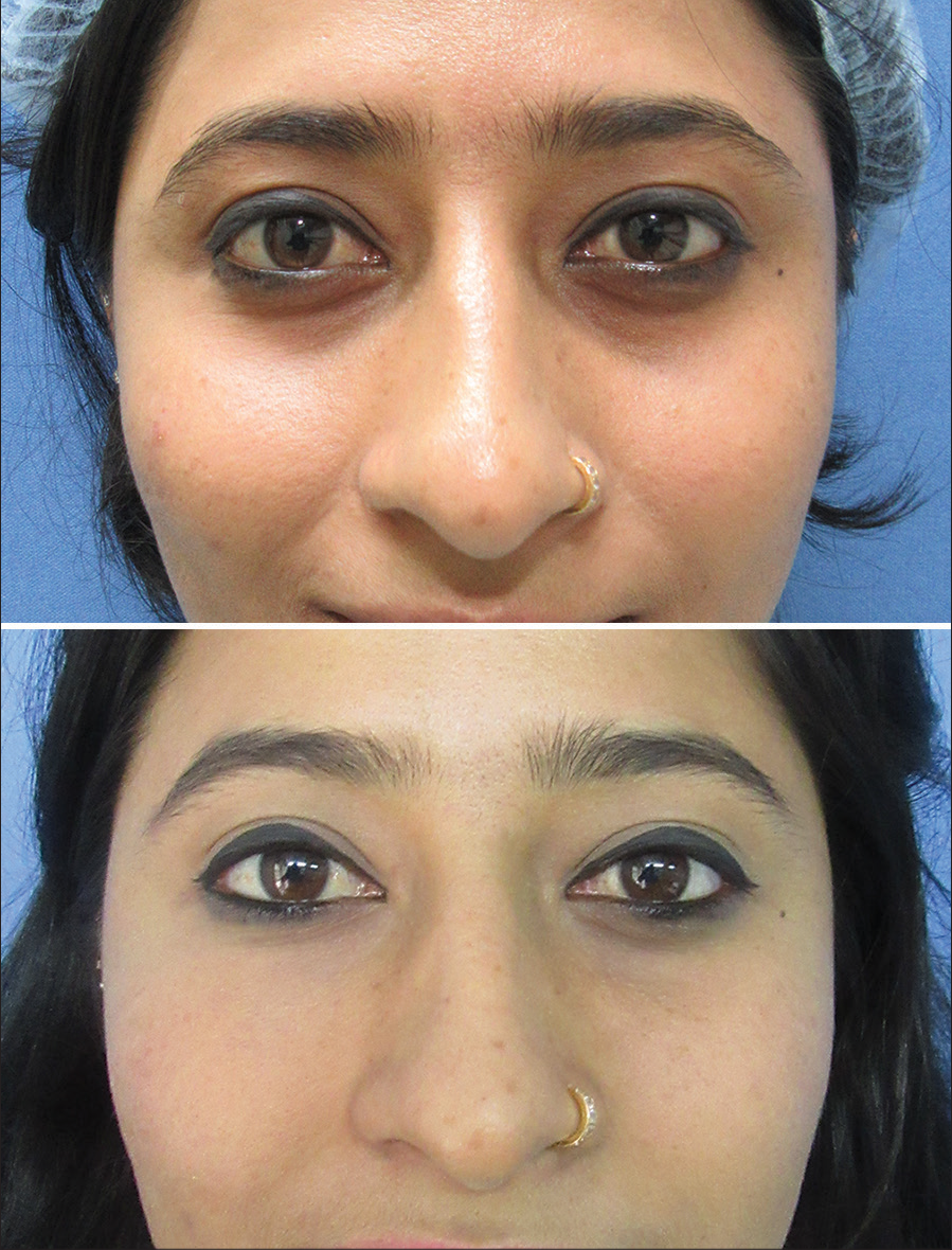
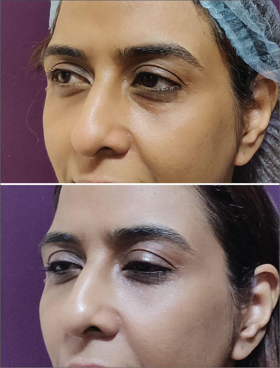
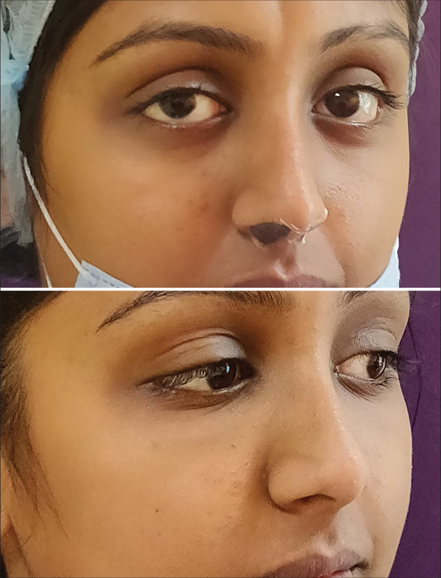
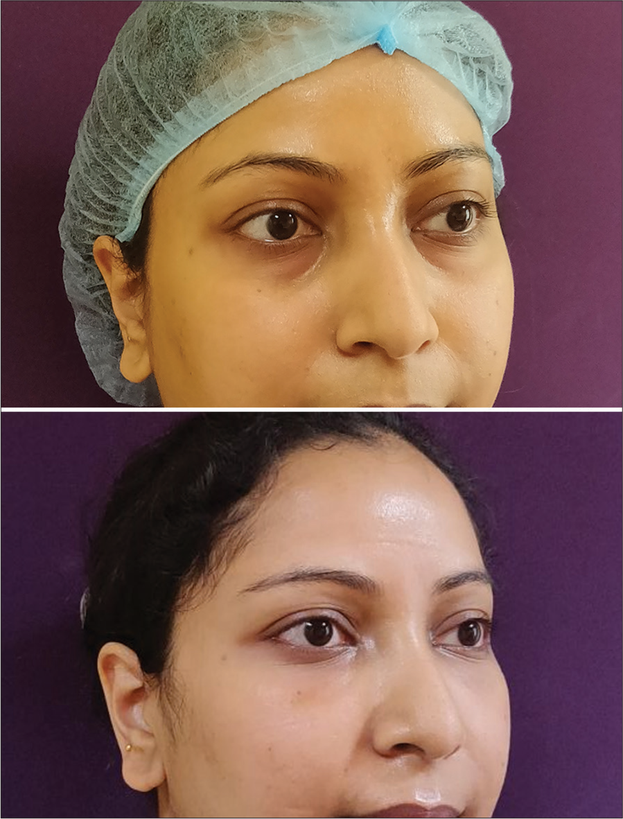
All patients were reviewed with Likert scoring at 3 months post-procedure. Out of the 36 patients, 28 (77%) rated their results as very satisfied (5), and 6 (17%) rated it as satisfied (4). One rated it as neutral (3) and 1 as unsatisfied (2). Both these patients underwent a second session of fat grafting where another 2 cc was injected per side. The overall Likert score was 4.7/5 [Figures 1-4].

- A 28-year-old patient with 4 cc microfat injected per side.

- A 40-year-old patient with 6 cc microfat injected per side.

- A 24-year-old patient with 5 cc microfat injected per side.

- A 32-year-old patient with 6 cc microfat injected per side.
Complications
Four patients (11%) experienced bruising, which resolved completely. All patients experience some edema, which resolves within 5 days. No other significant complications were reported.
DISCUSSION
Dark circles are a common complaint in young individuals.5 In an Indian study, it was found that periorbital melanosis was most prevalent in the age group of 16–25 years (i.e., 95 out of 200 patients [47.50%]). Among the 200 patients studied, it was more prevalent in women (162 [81%]) than men and the majority of the affected women were housewives (91 [45.50%]).3
Etiopathogenesis
Periorbital pigmentation has both genetic and lifestyle components, including lack of sleep, stress, alcohol overuse, and smoking.6,7 The pathogenesis of dark circles includes a lack of volume and hyperpigmentation. Pigmentation can be due to dermal melanocytosis,8 and stasis dyschromia due to poor venous drainage.9 Causes include atopic dermatitis, fixed drug eruptions, amyloidosis, lichen planus, and acanthosis nigricans. Volume deficiency can be due to anatomical orbital rim recession and age-related malar bone volume loss.10
Treatment
There exist a number of treatment options for dark circles. These can be divided into those targeting skin quality and those targeting volume.
Skin quality treatments include topicals, peels, and energy-based devices. Topical treatments aim at depigmentation by inhibiting tyrosinase activity, which converts Dihydroxyphenylalanine (DOPA) to melanin.11 The most commonly used agent is Hydroquinone. Other common agents include Kojic acid, Azelaic acid, Retinoic acid, Arbutin, and Vitamin C.12 Peeling agents remove melanin from the stratum corneum and epidermis, with deeper peels modulating melanin content in the dermis.13 However, they must be used carefully in darker skin-toned individuals as they can end up causing pigmentary complications.14 Commonly used peels include glycolic acid and trichloroacetic acid.
Lasers selectively target melanosomes while avoiding damage to surrounding structures.15,16 The most common ones are non-ablative Q-switched lasers such as ruby, alexandrite, and neodymium-doped yttrium aluminum garnet. Other lasers have also been tried such as pulsed dye, which targets hemoglobin,17 and ablative lasers such as carbon dioxide and erbium-doped yttrium aluminum garnet for resurfacing.18
In general, skin quality treatments need to be used for a significant period, usually months, before results are seen, and even then, they can be underwhelming. They can also be associated with side effects such as erythema, peeling, and burning.
Volume treatments
Hyaluronic acid fillers are the most popular treatment for volume replacement in the undereye area due to their ease, lack of downtime, rapid results, and reversibility. Numerous techniques have been described, including the use of needles, cannulas, deep bolus, and sandwiching techniques. In general, around 0.4–1 cc per side is required. Possible side effects include bruising, Tyndall effect, nodularity, and vascular complications. Hence, due caution needs to be exercised while performing the procedure.19-21
Fat grafting
While all the treatment options described thus far have varying degrees of efficacy, they are limited in being able to address only one component of the problem, that is, skin quality or volume loss. Hence, the combination of microfat and nanofat grafting is unique in being able to address both these issues in one procedure. As early as 1893, when Neuber used fat grafting for a depressed facial scar, he noticed an improvement in scarring along with depression.22 With the advent of liposuction, fat grafting increased in popularity, but early techniques used bolus techniques which had poor survival.23 In the 1990s, Coleman elucidated the process of separating, purifying, and injecting in small aliquots that enable more reliable results.24
With the increased popularity of fat grafting, multiple authors noted the improvement in the rejuvenation of overlying skin, presumably due to pluripotent adipose-derived stem cells (ADSC).25-27 The number of pluripotent cells contained in a cubic centimeter of adipose tissue is 100–1000 times larger than the number of stem cells contained in bone marrow.28 ADSCs also secrete various growth factors which have been shown to rejuvenate tissues.29-32 These growth factors include vascular endothelial growth factor, hepatocyte growth factor, insulin-like growth factor-1 and platelet-derived growth factor, keratinocyte growth factor, and fibroblast growth factor-1 and 2.
More pertinent to the treatment of dark circles, studies have shown that ADSCs can inhibit melanocyte proliferation and melanin synthesis. Interleukin 6 plays a key role in the downregulation of melanocytes.33
Stromal vascular fraction (SVF)
The SVF refers to all the cellular elements of the adipose tissue, excluding adipocytes. It includes smooth muscle cells, endothelial cells, blood cells, stem cells, and extracellular matrix which constitute 75% of the cell count of at tissue.34 The majority of the activity of SVF is attributable to ADSCs. However, evidence suggests that the presence of all the SVF cellular elements is helpful for ADSC activity due to the intercellular cross-talking.35
Two methods – chemical and mechanical help to obtain SVF. Chemical methods involve enzymatic digestion with collagenase, followed by filtration and centrifugation. This chemical SVF can then be used to culture ADCSs.36 Enzymatic techniques offer superior concentration but are limited by the additional time and expensive equipment required. Moreover, regulations in several countries pertaining to stem cells pose an additional hurdle.
Nanofat
Mechanical methods use physical disruption such as shaking, vortexing, and sonification, followed by washing and filtration. They contain fragments of extracellular matrix along with the cells.37 Mechanical SVF such as nanofat has found increasing popularity in the field of aesthetic regenerative therapy for the following reasons:
It is easier and cheaper to perform
It requires less equipment
It can be done in one sitting along with the harvest
It does not require any regulatory oversight.
Technique
While several methods have been described for autologous fat grafting to the face, we use the methods described by Tonnard et al. for preparing microfat and nanofat.38 The only way in which this study differs from the original description is by using sedimentation within the syringe itself as opposed to filtering through a nylon cloth. We prefer this method to be able to maintain a closed system where the fat is not exposed to the atmosphere at the point between harvest and injection, thereby reducing the risk of infection . Our infection rate of zero reinforces this technique. For the same reason, we also prefer sedimentation for fat separation and not centrifugation. A randomized comparison found no superiority for centrifugation for fat survival.39
Injection was done only in the supraperiosteal plane to avoid nodularity and visibility in this region. We also avoided parallel injections to avoid “sausaging” above the orbital ligament. Other authors have advocated this technique as well.40
Literature review
There is a paucity of literature on fat grafting for dark circles. Roh et al. published a series of 10 cases where fat was injected in the subcutaneous plane at serial intervals with cold storage. They reported a satisfaction rate of 78%.41 Youn et al. described using collagenase-digested fat cell grafts in 82 patients with improvement in 67% of cases.42 We were unable to find any study using nanofat for periorbital hyperpigmentation.
Complications
Swelling is usually present for a few days while bruising can occur on occasion. We did not have any incidence of infection. There have been several papers describing the safety of fat grafting for periorbital rejuvenation. One meta-analysis concluded that autologous fat grafting for the periorbital area provided a high satisfaction rate and did not result in severe complications. The five most commonly reported complications were edema, chemosis, contour irregularity deep wrinkles, and volume excess.43
Rare events of vascular occlusion have been reported as with fillers; hence, proper technique and knowledge of anatomy is essential.44
Limitations
Our paper is limited by being a case series. Moreover, there is a lack of standardization on fat preparation, volumes, and repeat procedures. We chose our techniques based on the “primum non nocere” principle. The other limitation is a lack of objective measurement of end points. To avoid any observer bias, we preferred patient self-evaluation.
CONCLUSION
Fat grafting and nanofat is an exciting treatment option for the treatment of dark circles, which is usually regarded as a recalcitrant problem. It has the advantages of addressing both volume and skin quality, being a single-stage procedure, and producing comparatively long-lasting results. At the same time, some downtime is expected when compared to other treatment options, and fat graft survival can be unpredictable on occasion. Proper technique is essential to avoid complications. But when done correctly, gratifying results can be obtained in a majority of cases. We hope our paper, which is the first to describe the use of nanofat for this condition, helps address the paucity of literature on fat grafting for dark circles. Author one conceptualized the paper, collected the data, performed the analysis, and prepared the paper.
Authors’ contributions
Author has conceptualized the paper, collected the data, performed the analysis, and prepared the paper.
Ethical approval
The Institutional Review Board approval is not required as this is a retrospective study.
Declaration of patient consent
The authors certify that they have obtained all appropriate patient consent.
Conflicts of interest
There are no conflicts of interest.
Use of artificial intelligence (AI)-assisted technology for manuscript preparation
The authors confirm that there was no use of artificial intelligence (AI)-assisted technology for assisting in the writing or editing of the manuscript and no images were manipulated using AI.
Financial support and sponsorship: Nil.
References
- Idiopathic cutaneous hyperchromia at the orbital region or periorbital hyperpigmentation. J Cutan Aesthet Surg. 2012;5:183-4.
- [CrossRef] [PubMed] [Google Scholar]
- What causes dark circles under the eyes? J Cosmet Dermatol. 2007;6:211-5.
- [CrossRef] [PubMed] [Google Scholar]
- Periorbital hyperpigmentation: A study of its prevalence, common causative factors and its association with personal habits and other disorders. Indian J Dermatol. 2014;59:151-7.
- [CrossRef] [PubMed] [Google Scholar]
- Infraorbital dark circles: Definition, causes, and treatment options. Dermatol Surg. 2009;35:1163-71.
- [CrossRef] [PubMed] [Google Scholar]
- International Society of Aesthetic Plastic Surgery. Available from: https://www.isaps.org/medical-professionals/isaps-global-statistics [Last accessed on 2024 May 01]
- [Google Scholar]
- Periorbital hyperpigmentation. An overlooked genetic disorder of pigmentation. Arch Dermatol. 1969;100:169-74.
- [CrossRef] [Google Scholar]
- Dark circles: Etiology and management options. Clin Plast Surg. 2015;42:33-50.
- [CrossRef] [PubMed] [Google Scholar]
- Condition known as “dark rings under the eyes” in the Japanese population is a kind of dermal melanocytosis which can be successfully treated by Q-switched ruby laser. Dermatol Surg. 2006;32:785-9.
- [CrossRef] [PubMed] [Google Scholar]
- Infraorbital Dark circles: A review of the pathogenesis, evaluation and treatment. J Cutan Aesthet Surg. 2016;9:65-72.
- [CrossRef] [PubMed] [Google Scholar]
- Relative maxillary retrusion as a natural consequence of aging: Combining skeletal and soft-tissue changes into an integrated model of midfacial aging. Plast Reconstr Surg. 1998;102:205-12.
- [CrossRef] [PubMed] [Google Scholar]
- Therapeutical approaches in melasma. Dermatol Clin. 2007;25:337-42, viii
- [CrossRef] [PubMed] [Google Scholar]
- Treatments of infra-orbital dark circles by various etiologies. Ann Dermatol. 2018;30:522-8.
- [CrossRef] [PubMed] [Google Scholar]
- Chemical peeling with trichloroacetic acid and lactic acid for infraorbital dark circles. J Cosmet Dermatol. 2013;12:204-9.
- [CrossRef] [PubMed] [Google Scholar]
- Periorbital hyperpigmentation: Review of etiology, medical evaluation, and aesthetic treatment. J Drugs Dermatol. 2014;13:472-82.
- [Google Scholar]
- Infraorbital pigmented skin. Preliminary observations of laser therapy. Dermatol Surg. 1995;21:767-70.
- [CrossRef] [PubMed] [Google Scholar]
- Combined therapy using Q-switched ruby laser and bleaching treatment with tretinoin and hydroquinone for periorbital skin hyperpigmentation in Asians. Plast Reconstr Surg. 2008;121:282-8.
- [CrossRef] [PubMed] [Google Scholar]
- Solar lentigines: Evaluating pulsed dye laser (PDL) as an effective treatment option. J Lasers Med Sci. 2013;4:33-8.
- [Google Scholar]
- Improvement of dermatochalasis and periorbital rhytides with a high-energy pulsed CO2 laser: A retrospective study. Dermatol Surg. 2004;30:483-7.
- [CrossRef] [PubMed] [Google Scholar]
- Use of hyaluronic acid filler for tear-trough rejuvenation as an alternative to lower eyelid surgery. Ophthalmic Plast Reconstr Surg. 2011;27:69-73.
- [CrossRef] [PubMed] [Google Scholar]
- Anatomic guidelines for augmentation of the cheek and infraorbital hollow. Dermatol Surg. 2012;38:1223-33.
- [CrossRef] [PubMed] [Google Scholar]
- Anatomical position of hyaluronic acid gel following injection to the infraorbital hollows. Ophthal Plast Reconstr Surg. 2013;29:35-9.
- [CrossRef] [Google Scholar]
- The fascinating history of fat grafting. J Craniofac Surg. 2013;244:1069-71.
- [CrossRef] [PubMed] [Google Scholar]
- The technique of periorbital lipoinflitration. Operat Tech Plast Reconstr Surg. 1994;1:120-6.
- [CrossRef] [Google Scholar]
- Clinical treatment of radiotherapy tissue damage by lipoaspirate transplant: A healing process mediated by adipose-derived adult stem cells. Plast Reconstr Surg. 2007;119:1409-22.
- [CrossRef] [PubMed] [Google Scholar]
- Adipose-derived stem cells for regenerative medicine. Circ Res. 2007;100:1249-60.
- [CrossRef] [PubMed] [Google Scholar]
- Basic science review on adipose tissue for clinicians. Plast Reconstr Surg. 2010;126:1936-46.
- [CrossRef] [PubMed] [Google Scholar]
- Adipose-derived stem and progenitor cells as fillers in plastic and reconstructive surgery. Plast Reconstr Surg. 2006;118:121S-8.
- [CrossRef] [PubMed] [Google Scholar]
- Secretion of angiogenic and antiapoptotic factors by human adipose stromal cells. Circulation. 2004;109:1292-8.
- [CrossRef] [PubMed] [Google Scholar]
- IFATS collection: Fibroblast growth factor-2-induced hepatocyte growth factor secretion by adipose-derived stromal cells inhibits postinjury fibrogenesis through a c-Jun N-terminal kinase-dependent mechanism. Stem Cells. 2009;27:238-49.
- [CrossRef] [PubMed] [Google Scholar]
- Hair growth promoting effects of adipose tissue-derived stem cells. J Dermatol Sci. 2010;57:134-7.
- [CrossRef] [PubMed] [Google Scholar]
- Adipocyte lineage cells contribute to the skin stem cell niche to drive hair cycling. Cell. 2011;146:761-71.
- [CrossRef] [PubMed] [Google Scholar]
- Adipose-derived stem cells inhibit epidermal melanocytes through an interleukin-6-mediated mechanism. Plast Reconstr Surg. 2014;134:470-80.
- [CrossRef] [PubMed] [Google Scholar]
- Understanding adipose-derived stromal vascular fraction (SVF) cell biology in reconstructive and regenerative applications on the basis of mononucleated cell components. J Prolother. 2013;10:15-29.
- [Google Scholar]
- Phenotypic analysis of stromal vascular fraction after mechanical shear reveals stress-induced progenitor populations. Plast Reconstr Surg. 2016;138:237e-47.
- [CrossRef] [PubMed] [Google Scholar]
- Enzymatic and non-enzymatic isolation systems for adipose tissue-derived cells: Current state of the art. Cell Regen. 2015;4:7.
- [CrossRef] [PubMed] [Google Scholar]
- Mechanical micronization of lipoaspirates: Squeeze and emulsification techniques. Plast Reconstr Surg. 2017;139:1369e-70.
- [CrossRef] [PubMed] [Google Scholar]
- Nanofat grafting: Basic research and clinical applications. Plast Reconstr Surg. 2013;132:1017-26.
- [CrossRef] [PubMed] [Google Scholar]
- A prospective, randomized comparison of clinical outcomes with different processing techniques in autologous fat grafting. Plast Reconstr Surg. 2022;150:955-62.
- [CrossRef] [PubMed] [Google Scholar]
- Treatment of infraorbital dark circles by autologous fat transplantation: A pilot study. Br J Dermatol. 2009;160:1022-5.
- [CrossRef] [PubMed] [Google Scholar]
- Correction of infraorbital dark circles using collagenase-digested fat cell grafts. Dermatol Surg. 2013;39:766-72.
- [CrossRef] [PubMed] [Google Scholar]
- Efficacy, safety and complications of autologous fat grafting to the eyelids and periorbital area: A systematic review and meta-analysis. PLoS One. 2021;16:e0248505.
- [CrossRef] [PubMed] [Google Scholar]
- Ophthalmic complications following facial autologous fat graft injection: A systematic review and meta-analysis. Aesthetic Plast Surg. 2022;46:3013-35.
- [CrossRef] [PubMed] [Google Scholar]


