Translate this page into:
Postauricular Pull-through Transposition Flap for an Anterior Auricular Defect
Address for correspondence: Dr André Cerejeira, Department of Dermatology and Venereology, Centro Hospitalar São João, Alameda Prof. Hernâni Monteiro, 4200-319 Porto, Portugal. E-mail: cerejeira.andre@gmail.com
This is an open access journal, and articles are distributed under the terms of the Creative Commons Attribution-NonCommercial-ShareAlike 4.0 License, which allows others to remix, tweak, and build upon the work non-commercially, as long as appropriate credit is given and the new creations are licensed under the identical terms.
This article was originally published by Wolters Kluwer - Medknow and was migrated to Scientific Scholar after the change of Publisher.
Dear Editor,
Defects involving the anterior part of the ear are relatively common in dermatological surgery, especially after tumor ablation. The complexity and tissue characteristics of this anatomical area render reconstructive procedures challenging.
The postauricular pull-through transposition flap is a versatile reconstructive option that allows a one-stage reconstruction, usually with a good cosmetic result.[1] Advantages of this flap also include the possibility to cover large defects and the usage of well-vascularized and protected skin.[2]
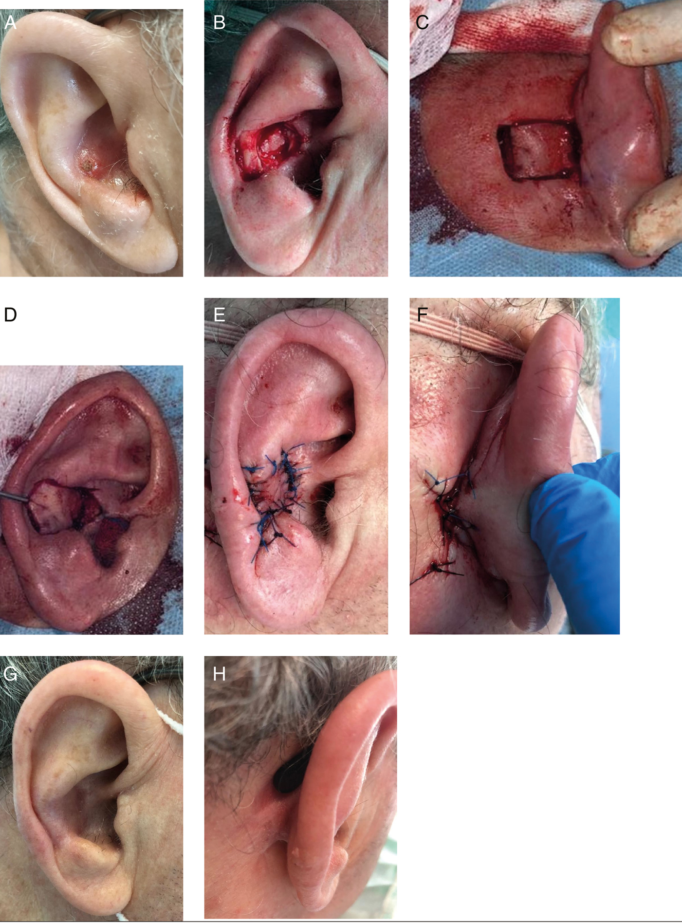
A 67-year-old man, with no significant medical history, presented with a squamous cell carcinoma of the right conchal bowl and antihelix [Figure 1A]. Tumor extirpation was through the dermis and perichondrium, exposing intact auricular cartilage. The tumor and a margin of 4 mm were excised, resulting in a 2.4 × 1.7 cm defect [Figure 1B]. Margins free of disease were confirmed by extemporaneous histopathological examination. The auricle was reflected anteriorly, and an area of donor skin was measured and marked just posterior to the postauricular sulcus. The flap was then peripherally excised and meticulously undermined, preserving a subcutaneous pedicle that originated from the postauricular sulcus [Figure 1C]. Returning the auricle to its normal anatomical position, a tunnel was created connecting the base of flap’s pedicle to the median margin of the defect. The flap was then pulled and adapted to the defect, without tension, torsion, or impingement of the pedicle [Figure 1D]. The flap was inset with fine nonabsorbable sutures [Figure 1E], and the secondary defect was easily closed primarily [Figure 1F]. A standard pressure dressing was applied. No postoperative complications were noted. Postoperative antibiotic prophylaxis was performed using ciprofloxacin. Nine months after surgery, an excellent cosmetic outcome was observed [Figure 1G and H].

- A: Squamous cell carcinoma of the right conchal bowl and antihelix. B: Defect of the right conchal bowl and antihelix measuring 2.4 × 1.7 cm. C: Flap donor site was marked and peripherally excised, preserving a subcutaneous pedicle that originated from the postauricular sulcus. D: The flap was then pulled through a tunnel and adapted to the defect, without tension, torsion, or impingement of the pedicle. E: The flap was sutured into place. F: The secondary defect was easily closed primarily. G: Nine months after surgery, an excellent cosmetic outcome was observed (lateral view). H: Nine months after surgery, an excellent cosmetic outcome was observed (posterior view)
Various reconstructive procedures were considered to manage the described surgical defect. Primary closure was not an option as it could originate distortion of the auricular anatomy. As the cartilage was exposed, a skin graft could be associated with a high risk of necrosis. Second intention healing would be a long process with a significant risk of infection. Two-stage local flaps transposed from the postauricular or preauricular regions require a second stage to remove the pedicle.
Originally described by Masson,[3] the postauricular pull-through transposition flap is appropriate for both small and large defects of the scaphoid fossa, antihelix, and conchal bowl. The postauricular skin offers safe vascularization and has similar color and texture when compared with the auricular skin. Furthermore, the resulting scars remain well hidden.
The main risks of this flap are infection and necrosis. The latter is related primarily with the dimension of the cartilaginous opening that allows passage of the flap, which must be wide enough.[2] Vascular supply from tributaries of the posterior auricular artery contributes to the viability of this flap. In our case, neither infection nor necrosis was observed.
In conclusion, the pull-through postauricular transpositional flap is a safe and relatively simple one-stage flap associated with satisfactory functional and aesthetic outcomes and minimal morbidity. It is an option worth considering in the reconstruction of defects of the anterior auricle.
Declaration of patient consent
The authors certify that they have obtained all appropriate patient consent forms. In the form the patient(s) has/have given his/her/their consent for his/her/their images and other clinical information to be reported in the journal. The patients understand that their names and initials will not be published and due efforts will be made to conceal their identity, but anonymity cannot be guaranteed.
Financial support and sponsorship
Nil.
Conflicts of interest
There are no conflicts of interest.
REFERENCES
- Postauricular flap based on a dermal pedicle for ear reconstruction. Plast Reconstr Surg. 1981;68:159-65.
- [Google Scholar]
- Anterior conchal reconstruction using a posteroauricular pull-through transpositional flap. Plast Reconstr Surg. 2004;113:2071-5.
- [Google Scholar]
- A simple island flap for reconstruction of concha-helix defects. Br J Plast Surg. 1972;25:399-403.
- [Google Scholar]


