Translate this page into:
Two-stage Ear Reconstruction with a Retroauricular Skin Flap after Excision of Trichilemmal Carcinoma
Address for correspondence: Dr. Shashank Bhargava, 32, Varruchi Marg, Madhav Nagar, Freeganj Ujjain- (MP) 456010, India. E-mail: shashank2811@gmail.com
This is an open access journal, and articles are distributed under the terms of the Creative Commons Attribution-NonCommercial-ShareAlike 4.0 License, which allows others to remix, tweak, and build upon the work non-commercially, as long as appropriate credit is given and the new creations are licensed under the identical terms.
This article was originally published by Wolters Kluwer - Medknow and was migrated to Scientific Scholar after the change of Publisher.
Abstract
Abstract
Trichilemmal carcinoma is a rare cutaneous tumor with a frequently good prognosis but without standard criteria for surgical treatment. We aimed to show the results of a two-stage surgical approach that preserves the anatomical features of the ear. We report a case of 82-year-old man with trichilemmal carcinoma of the ear that was treated with a two-stage surgical approach. We observed that 5 months after the surgeries, the ear appeared entirely healed and there were no signs of recurrence; hence, our two-stage surgical approach allowed the anatomy of the ear to be preserved after the complete excision of the tumor.
Keywords
Trichilemmal carcinoma
two-stage surgical approach
retroauricular skin flap
ear reconstruction
INTRODUCTION
The trichilemmal carcinoma (TC) is a very uncommon cutaneous adnexal tumor that typically occurs on sun-exposed skin, mainly on the face, scalp, and neck regions.[1] It is a malignant neoplasm with an indolent clinical course that originates from the outer root sheath of the hair follicle.[2] It usually affects the elderly patients due to cumulative solar radiation exposure, and no sex predilection is present. In our case, TC presented in the posterior part of right helix, an area that has only been described once in the available medical literature.[2]
CASE REPORT
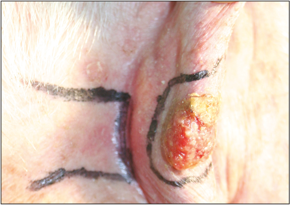
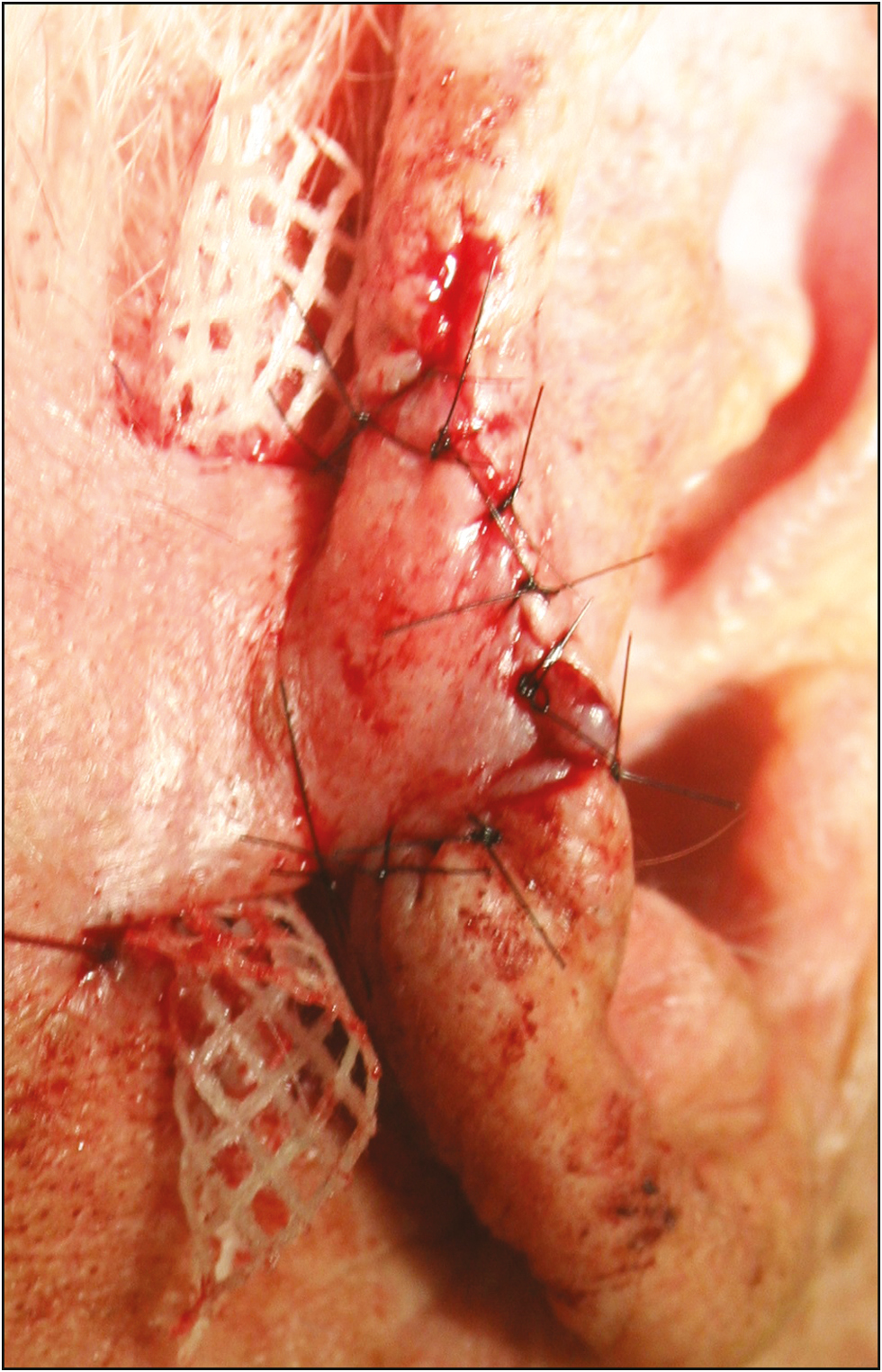
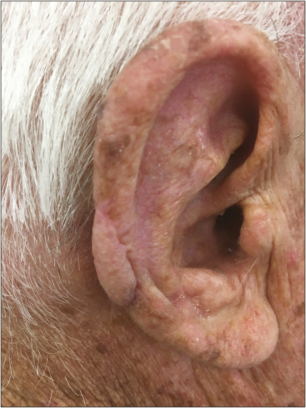
An 82-year-old man presented to our hospital in October 2017, with a 2 × 1.5cm nodular ulcerated, crusted, and mildly painful lesion of the posterior part of the auricle. The lesion had appeared approximately 1 year earlier and it had never been treated. The patient was a phototype II with blue eyes, and his skin was remarkably photodamaged with several actinic keratoses. We aimed to achieve a complete excision of the tumor with a good functional and aesthetic outcome, and we opted for a two-stage approach. The first stage consisted of wide local excision of the lesion of the helix [Figure 1]. The resulting tissue defect was closed with a trapezoid-shaped skin flap, approximately 3.5 × 2cm, which was advanced from the mastoid region and attached with 5-0 nylon sutures. The mastoid’s loss of tissue was covered with a full-thickness skin graft from supraclavicular region, which was also sutured with a 5-0 Vicryl rapid. The donor area of the skin graft was sutured with 4-0 nylon. After 3 weeks, the auricular flap appeared well perfused and ready for a second operation to detach the skin flap from the mastoid donor area, and to shape the new ear [Figure 2]. The anatomopathological examination of the excised specimen revealed an ulcerated neoplasm, characterized by histological aspect compatible with TC. Ultrasound imaging was performed, which did not identify signs of metastasis or deep invasion. Five months after the operation, the ear appeared entirely healed, and no signs of recurrence were observed. The mastoid donor site had completely healed as well [Figure 3].

- The first stage consisted of wide local excision of the lesion of the helix, the incision markings can be seen around the tumor

- After second stage of the operation to detach the skin flap from the mastoid donor area and to shape the new ear

- Five months after the operation, the ear appeared entirely healed, and no signs of recurrence were observed
DISCUSSION
The term “TC” was first introduced by Headington.[3] It is considered the malignant counterpart of trichilemmoma with low metastasis potential and usually good prognosis.[45] Nevertheless, seldom cases of deep invasion and local recurrence have been reported in the literature.[6] Furthermore, in immunosuppressed transplanted patients, TC can have a very aggressive evolution with the development of metastasis to the liver and the lung. The pathogenesis of TC is not completely clear but sun exposure seems to be the major causative agent. However, this tumor has also been reported after radiotherapy treatment and it seems that preexisting burn scars may also predispose to its development. No standard treatment is available for TC. Surgical excision with 1cm safety free margins is considered the first choice for curative treatment, and regular surveillance without any adjuvant therapy is generally sufficient.[7] Mohs micrographic surgery can be performed to track the pagetoid spread that sometimes is far into clinically normal skin.[2] We have chosen to perform an intervention that would allow the anatomy of the ear to be preserved in its size, shape, and fold of the helix, without renouncing the safety of the procedure with regard to the complete excision of the tumor. The flap that we used allowed us to reconstruct the helix profile, restoring the natural anatomical thickness. Finally, this two-staged approach can be performed in the same day clinic under local anesthesia, avoiding the risks and complications of a general anesthesia, which becomes even more relevant in consideration of the generally advanced age of the patients.
CONCLUSION
Our two-stage surgical approach allowed the anatomy of the ear to be preserved after the complete excision of the tumor.
Declaration of patient consent
The authors certify that they have obtained all appropriate patient consent forms. In the form the patient(s) has/have given his/her/their consent for his/her/their images and other clinical information to be reported in the journal. The patients understand that their names and initials will not be published and due efforts will be made to conceal their identity, but anonymity cannot be guaranteed.
Financial support and sponsorship
Nil.
Conflicts of interest
There are no conflicts of interest.
REFERENCES
- Folliculocentric squamous cell carcinoma with tricholemmal differentiation: a reappraisal of tricholemmal carcinoma. Clin Exp Dermatol. 2012;37:484-91.
- [Google Scholar]
- Recurrent and metastatic trichilemmal carcinoma of the skin over the thigh: a case report. Cancer Res Treat. 2010;42:176-9.
- [Google Scholar]


